The Department of Radiology and Imaging is nationally and internationally recognized as the premier center for leading-edge musculoskeletal, orthopedic and rheumatologic clinical and research imaging. Our mission is to provide the highest quality diagnostic imaging for musculoskeletal conditions and to provide image-guided treatment options to support restoration of function and mobility. Our goal is to enhance the quality of patient lives through cutting-edge research in diagnostic imaging – in MRI, CT, ultrasound and interventional radiology – through the development of new techniques that optimize the early detection and treatment of musculoskeletal conditions.
For information regarding insurance participation status for all HSS Radiologists, please call 212.774.2607.


The Department of Radiology & Imaging facilities include a wide array of state-of-the-art imaging technologies to provide patients with clear and accurate results. Our highly experienced staff is available to answer any questions or give further explanation of our services.
Below is a list of the services offered:
Copies of the reports from your imaging examination can be obtained through your referring physician's office. The physician can call our Image Records Department at 212.606.1135. Reports can be faxed or mailed free of charge to their office. There is a $35 charge per CD for copies of your images which can hold several imaging examinations, or $12 per sheet of X-ray film. Please have your name, address, date of birth, date of service, and type of exam readily available.
For your protection, patients requesting images and/or reports are required to complete a Request for Release of Information and fax it to 212.774.2327. Patients may pick up reports and/or images personally with proper ID for verification at The Belaire Building Image Records Department located at 525 East 71st Street, Suite 4K, New York, NY 10021. We also offer free FedEx ground shipping. For additional information please call 212.606.1135.
The Department of Radiology and Imaging provides safety and security of all patients, employees and visitors in regard to medical imaging including radiation hygiene, interventional procedures performed under imaging guidance and the use of contrast agents for enhanced imaging outcomes. Administrative controls and oversight are in place to establish policies and procedures which assure safety and security.
Members of the Department of Radiology and Imaging are musculoskeletal-fellowship-trained, board certified radiologists specializing in the interpretation of radiological tests and diagnosis of disease. The bill from HSS Radiologists is for the personal professional consultative services of the individual radiologist.
The separate, Hospital bill is for technical charges to cover the costs of the equipment, supplies, and services performed by the radiology technicians and other Hospital personnel in regard to the radiological tests performed.
What your bills for radiology services represent:
Radiologist Bill
Charges cover radiologist consultations on one more of the following:
Hospital Bill
Charges cover:
Questions regarding your bill
For questions regarding the HSS Radiologists bill call 866.689.8865
For questions regarding the Hospital bill call:
Inpatient Services:
Last name starting with A - L: 212.606.1388
Last name starting with M - Z: 212.606.1034
Outpatient Services: 212.606.1772
Billing Information
If you have insurance coverage from Medicare, Medical Assistance, a participating managed care carrier or a commercial healthcare insurer, and you have provided complete financial information to the Hospital at the time you received services, we will submit a claim to your carrier on your behalf.
Radiologic Interpretation
All imaging studies performed at HSS are interpreted by a musculoskeletal-fellowship-trained, board certified radiologists with subspecialty experience in the manifestations of orthopedic and rheumatic conditions. Board certification in radiology requires years of training beyond medical school, internship, residency, fellowship, as well as the satisfaction of both written and oral examinations. Certification by the American Board of Radiology assures quality care, expertise in quality image acquisition and safe use of ionizing radiation and magnetic resonance.
Official interpretation by an experienced radiologist enables subtle X-ray findings (for example, infections, systematic diseases, tumors, fracture, etc.) to be detected early, before an abnormality has progressed to where it is more obvious and easier to recognize from the imaging. A delay in getting an accurate diagnosis can considerably, negatively affect treatment and, ultimately, patient outcomes. It also adds to patient discomfort, inconvenience and increased medical costs. This is why early, expert interpretation of imaging by an experienced radiologist is key.
Quality of care is top priority at HSS. In order to assure quality of care and patient protection, HSS follows The Joint Commission (TJC) in requiring an official interpretation and an authenticated report by a radiologist for all imaging procedures and examinations performed in an accredited hospital. HSS is an accredited hospital and official interpretations of imaging studies are conducted by board certified radiologists with subspecialty training. Images generated and acquired by HSS, and the reports on their interpretation, are maintained in the Hospital medical records.
The Board of Trustees, the HSS administration, and the physicians of HSS are dedicated to delivering excellent quality patient care but are also concerned with escalating medical costs. Keeping costs down while assuring that cost-containing restrictions do not jeopardize a person's health, well-being or access to quality care is the responsibility of every physician. The radiologists and the staff of the Department of Radiology and Imaging take pride in reviewing orders for imaging examinations so as to eliminate unnecessary examinations and to prevent redundancy.
HSS Main Campus (MRI, CT, US, Fluoroscopy, X-ray imaging)
535 East 70th Street
New York, NY 10021
75th Street Campus (MRI, X-ray imaging, Bone Density)
429 East 75th Street
New York, NY 10021
HSS West Side (MRI, CT, US, X-ray imaging)
610 W 58th Street
New York, NY 10019
HSS Hudson Yards (X-ray imaging)
31 Hudson Yards
10th Floor
New York, NY 10001
HSS ASC of Manhattan (X-ray imaging)
1233 Second Avenue
New York, NY 10065
HSS Brooklyn (MRI, X-ray imaging)
148 39th Street
7th Floor
Brooklyn, NY 11232
HSS Queens (X-ray imaging)
176-60 Union Turnpike
Suite 190
Fresh Meadows, NY 11366
HSS Long Island (MRI, X-ray imaging)
333 Earle Ovington Blvd.
Suite 106
Uniondale, NY 11553
HSS Stamford (MRI, X-ray imaging, Ultrasound)
1 Blachley Road
Stamford, CT 06902
HSS Southampton (X-ray Imaging)
56 Flying Point Road, Unit 4
Southampton, NY 11968
HSS Paramus (MRI, X-ray imaging)
140 East Ridgewood Avenue
Suite 175 S.
Paramus, NJ 07652
HSS Westchester (MRI, X-ray imaging)
1133 Westchester Avenue
White Plains, NY 10605
HSS Florida (MRI, X-ray imaging)
300 Palm Beach Lakes Blvd
West Palm Beach, FL 33401

BA: Vanderbilt University
MD: University of Kentucky College of Medicine
Internship: Naval Medical Center San Diego
Residency: Icahn School of Medicine at Mount Sinai

BS: Williams College
MD: Columbia University Vagelos College of Physicians and Surgeons
Internship: Englewood Hospital
Residency: New York-Presbyterian Hospital/Weill Cornell Medical College

BS/MS: Northwestern University
MD: Tufts University School of Medicine
Internship: Flushing Hospital
Residency: Stony Brook University Hospital

MD: Tehran University of Medical Sciences
Internship: St. Agnes Hospital
Residency: Yale School of Medicine
Class of 2024
Class of 2023
Class of 2022
Class of 2021
Class of 2020
Class of 2019
Class of 2018
Class of 2017
Class of 2016
Class of 2015
Class of 2014
Class of 2013
The fellows enjoy a robust conference schedule to round out their education. Protected time is given for daily radiology conferences. Our radiology attending staff regularly attend (virtually and/or in-person) and participate in every conference. Oftentimes, there are more than 10 radiology attendings at conference, with each adding his or her perspective on the presented topic. Conferences last at least one hour.
Weekly Radiology Conferences:
Additional conferences that fellows regularly attend:
Research is a vital component of fellowship training within the department of Radiology at HSS. A wide variety of research opportunities are available to Musculoskeletal Radiology Fellows, encompassing a spectrum of research designs ranging from simple case reports to NIH funded long term prospective studies. Projects pertaining to every imaging modality performed at HSS are available to fellows, as are collaborative projects with other hospital services and outside institutions. HSS Radiology attendings are available as research mentors should a fellow wish to pursue his or her own research ideas.
A requirement of the fellowship is submission of at least one abstract for consideration for the end of year HSS Clinical Fellow Research Presentations. Fellows will be required to select at least one project of sufficient scientific merit (i.e. data/hypothesis driven) in order to fulfill this requirement.
Research submitted to major national and international conferences is encouraged and reimbursed.
HSS MRI Lab: The HSS MRI lab is a multidisciplinary group of individuals, including radiologist clinician scientists, biomedical engineers, MRI physicists, veterinary radiologists, and computational scientists whose collaborative efforts are aimed at furthering advanced MR imaging techniques and their translation to clinical applications. Current research topics include MR imaging in the presence of metallic implants, parametric imaging of musculoskeletal tissues, advanced neurography techniques, and AI based image reconstruction. Ongoing research projects available to fellows within the MRI lab include investigation of MRI as a biomarker for adverse local tissue reaction in total hip arthroplasty, T2* relaxometry of the ACL in pre- and post-menopausal females, and AI based image reconstruction for 2D/3D cervical spine MRI, among many others.
Ultrasound Research: Research opportunities within the division of ultrasound are plentiful and include a number of grant-funded studies on a variety of topics. Current ultrasounds research topics include quantitative imaging of muscle and tendons, advanced neurosonography, and fracture healing. Ongoing research projects available to fellows within the ultrasound division include ultrasound of tendon impingement in the setting of distal radial fractures, ultrasound/MRI correlation of synovitis in total knee arthroplasty, and ultrasound of anatomic variations of the brachial plexus.
Research opportunities pertaining to interventional radiology, computed tomography, and radiographic imaging are also available. Current fellow projects include utility of preoperative chest radiographs in the COVID era, C1-2 interventions under CT and fluoroscopic guidance, and effects of music therapy during spinal injections.
Fellows will take call for one week at a time beginning Friday evening and ending the following Friday morning. An attending is scheduled and readily available on back-up call for consultation. An on call laptop is provided and each fellow is given a monitor for taking call at home.
Stipend: $96,976 (2020–2021) per annum/benefits (subsidized housing also available)
Vacation and Educational/Conference Time: Each fellow is entitled to four weeks of paid vacation time (20 working days). Additional time for conference attendance may be granted based on factors including appropriateness and value of workshop, conference, or seminar to the Fellow’s training and responsibilities, whether the Fellow is presenting research findings at the conference, as well as staffing needs.
Other benefits:
As a dedicated musculoskeletal imaging facility, Hospital for Special Surgery's Department of Radiology and Imaging focuses all research, training, and the development of new techniques on optimizing the ability to image the earliest signs of a musculoskeletal condition, disease progression and/or healing.
HSS performs novel MRI research to aid in the early diagnosis of orthopedic conditions, the surgical planning, and the long term evaluation of patients with orthopedic diseases.
Learn more about the MRI Research Lab.
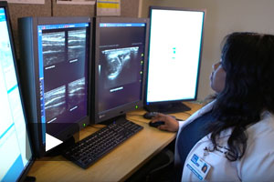
The Division of Ultrasound at HSS boasts a five-room patient examination suite dedicated to musculoskeletal ultrasound. All ultrasound systems are equipped with high-resolution transducers and have capability to perform both strain (qualitative) and shear wave (quantitative) elastography. Our radiologists are principal investigators and co-investigators on numerous grant-funded research efforts originating within the Division of Ultrasound, and are well-published in the field of musculoskeletal ultrasound. Interdepartmental collaborations at HSS have also expanded musculoskeletal ultrasound research into orthopedics, biomechanics and rheumatology. Current grant-funded ultrasound research topics include neurosonology, quantitative muscle and tendon imaging, and fracture healing.