MRI (Magnetic Resonance Imaging)
Magnetic resonance imaging (MRI) is a diagnostic imaging technology used to detect problems in both bone and soft tissues.
MRI depicts soft-tissue injury and abnormalities with greater sensitivity and specificity than conventional imaging techniques. Hospital for Special Surgery uses high-resolution MRI to demonstrate fine details of articular cartilage, tendons, peripheral nerve and other soft-tissue structures that are not always demonstrated on routine MRIs used elsewhere.
Below are frequently asked questions about MRIs and their answers.
Why has my doctor ordered an MRI exam?
Most people want to know why they are having symptoms of a physical problem. Your doctor has ordered an MRI to make, confirm or exclude a diagnosis with treatment of your condition as the goal.
Schedule an MRI exam at HSS
(A prescription is required)
Who performs and interprets the MRI exam?
Your exam will be performed by a technologist who has years of training in specialized magnetic resonance imaging, under the direction of an attending radiologist. The attending musculoskeletal radiologist, who specializes in and has advanced training in orthopedic MRI, will protocol the examination of the bones and soft tissues in the area of interest and interpret your examination.
A radiologist is a doctor specializing in all imaging modalities including MRI, ultrasound and CT scans. Radiologists specialize in the imaging and diagnosis of disease. Interpretation of a radiograph MRI, CT or ultrasound examination requires expertise in pattern recognition and in the identification of potential artifacts that may otherwise be mistaken for pathology. Radiologists are trained in the variable sensitivity and specificity of each imaging technique, and in the potential for hazards related to the examination that could cause harm and must be avoided.
All the radiologists at Hospital for Special Surgery are board certified. Having years of experience in the imaging of musculoskeletal disorders, and the majority have additional formal fellowship training beyond residency in musculoskeletal or body imaging.
How is an MRI performed?
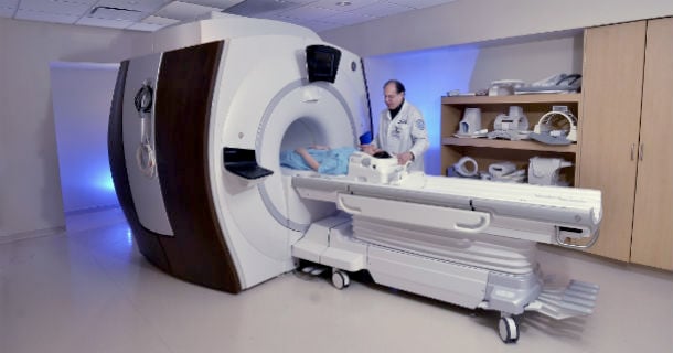
There is little or no risk in having an MRI exam. You will lie on a table within a high strength external magnet. During the examination, you will hear loud banging which is electrical gradients that drive the machine.
What should I do to prepare for an MRI?
Persons with severe claustrophobia may consider taking a sedative with their doctor's approval. If you are a pregnant woman in the first trimester, an MRI examination is not recommended. After that, you should obtain the approval of your obstetrician.
Preparing for an MRI exam is easy. Unless your doctor tells you otherwise, you may take your medications as usual. There are no food or drink restrictions either. The only unusual preparation for an MRI scan is that all removable metallic objects must be left outside the scanning room. These include:
- jewelry
- keys
- watches
- coins
- eyeglasses
- hearing aids
- dentures and other prosthetic devices
Credit cards should not be brought anywhere near the MRI magnet. Since they are magnetically coded, the MRI’s magnet, which is very powerful, can easily corrupt the information stored on them. You will be asked to change into a hospital gown.
What are the risks in an MRI?
MRI is one of the safest diagnostic exams available. Unlike X-rays and computerized tomography (CT), MRI does not use radiation. However, if you wear a pacemaker or have certain body implants (such as cochlear ear implants), you should not have an MRI examination. You will fill out a questionnaire prior to the MRI to ensure your absolute safety.
What if I need an MRI with IV contrast?
Your referring provider may order an MRI with an IV gadolinium-based contrast agent to better help see problems. At HSS, Gadavist is used. This medication is manufactured by Bayer HealthCare Pharmaceuticals Inc.
- What is Gadavist?
- Gadavist is a gadolinium-based contrast agent (GBCA) used in MRI that is available through prescription. Gadavist, like other GBCAs, is injected into your vein during some MRI studies. It is considered one of the safest GBCA contrast agents available.
- What are the benefits of Gadavist and why do we use Gadavist?
- Gadavist is a safe contrast agent that helps radiologists diagnose certain diseases by enhancing the MRI images. Prior to requesting Gadavist for your MRI, your doctor will review your medical records to determine the safety and benefit of using Gadavist for your MRI exam.
- What are the risks of Gadavist?
- All medications come with risks. For Gadavist, allergic reaction is one of them.
- Though rare, the most common allergic reactions to Gadavist are headache, nausea, dizziness, and rash. Serious complications like nephrogenic systemic fibrosis (NFS) are also extremely rare but can occur in patients with kidney failure. Appropriate screening/assessment is done in advance to avoid these reactions.
- Can Gadavist stay in your body after MRI procedure?
- Gadavist contains a metal called gadolinium. Small amounts of gadolinium can stay in your body for several months to years. It is not known how gadolinium retention may affect you in the future, but so far, studies have not found harmful effects in patients, especially those with normal kidneys.
- What are the ingredients in Gadavist?
- Active ingredient: gadobutrol
- Inactive ingredients: calcobutrol sodium, trometamol, hydrochloric(for pH adjustment), and water for injection.
What are the alternatives to having an MRI?
Alternative tests to MRIs can include CT scans or ultrasound imaging, both of which are non-invasive. A CT (computed tomography) scan uses high-speed computers and a series of thin X-ray beams to obtain 360-degree X-ray view of a selected area of the body. A musculoskeletal ultrasound uses sound waves instead of ionizing radiation to produce images of soft tissues.
What can I expect after the MRI examination?
There are no after effects of an MRI examination. Following an MRI examination, you will immediately be able to resume your pre-examination activities.
What happens with my MRI results?
The radiologist will generate a written report, which will be available to your physician within 24 to 48 hours of the MRI exam. If your physician is at HSS, the images will be available immediately following the exam. The resulting report is sent to your referring physician and will become part of the permanent record. Your physician will review the MRI test results with you and can integrate the results of your MRI test with the findings on your physical examination and laboratory tests.
Copies of the report can be obtained through your referring physician's office. Your physician can call the file room at 212.606.1135 and a copy of the report can be faxed or mailed, free of charge, to their office. The images are the property of the institution, as are biopsy slides or blood samples. Copies of the images can be obtained on a CD by contacting the file room. There is a charge for obtaining CDs and mailing them to your physician.
Will other tests be ordered?
Additional tests to assess your problem may be ordered before or after the MRI at the discretion of your doctor.
What is special about MRIs at HSS?
Hospital for Special Surgery uses high-resolution MRIs that show details in soft tissues that are not always visible on the MRIs used routinely at many other healthcare organizations. We operate the largest academic orthopedics-dedicated MRI facility in the world, with more than 10 MRI units of different field strengths, enabling HSS to meet the diagnostic needs of a greater number of patients with a wider variety of conditions. All HSS radiologists at are board certified with years of experience in musculoskeletal imaging and advanced pattern-recognition expertise to identify potential artifacts that may otherwise be mistaken for an injury or disease. At HSS:
- Dedicated coils are used for specific body parts to obtain the best images.
- MRIs are performed utilizing high-matrix imaging/thin-sliced imaging.
- Examinations of joints are performed without intra-articular injections. These are highly diagnostic, non-invasive scans.
- Techniques HSS have been validated for accuracy and published in peer-reviewed research.
- Customized orthopedic protocols have been developed specific to each referring physician's specialized surgical needs.
- Specialized scanning is performed for detailed diagnostic examinations of patients who have orthopedic hardware and total joints implants.
- Ultra-wide short bore units available to benefiting patients of all sizes, as well as those patients with a history of severe claustrophobia.
- The Division of MRI is a GE Luminary site for testing coils and protocols for optimal musculoskeletal imaging.
Can I have an MRI if I have a hip or knee replacement implant?
At Hospital for Special Surgery, you can. The MRI division of the HSS Department of Radiology has safe and accurate ways to do MRIs to assess potential causes of pain after a joint replacement, such as implant loosening and adverse tissue reactions, as well as for unrelated conditions in the same part of the body, such as including tendonitis or muscle tears. Our doctors and scientists have published substantial research in this area and, through our Magnetic Resonance Imaging Lab, continually work together to develop new techniques that overcome the artifacts imposed by imaging around metallic implants. This greatly enhances the quality of imaging and enables more accurate diagnoses.
References
- Alamanda VK, Demartino I, Potter HG, Koff MF, Lin B, Muskat A, Westrich GH. Multiacquisition Variable-Resonance Image Combination Magnetic Resonance Imaging Used to Study Detailed Bone Apposition and Fixation of an Additively Manufactured Cementless Acetabular Shell. Arthroplast Today. 2020;6(4):694-698. Published 2020 Aug 26. doi:10.1016/j.artd.2020.07.019.
- Burge AJ, Jawetz ST. Advanced Magnetic Resonance Imaging in Osteoarthritis. Semin Musculoskelet Radiol. 2020;24(4):355-366. doi:10.1055/s-0040-1708822.
- Koff MF, Burge AJ, Potter HG. Clinical magnetic resonance imaging of arthroplasty at 1.5 T. J Orthop Res. 2020;38(7):1455-1464. doi:10.1002/jor.24606.
- Lee DH, Choi YS, Potter HG, Endo Y, Sivakumaran T, Lim TK, Chun TJ. Reverse total shoulder arthroplasty: an imaging overview. Skeletal Radiol. 2020;49(1):19-30. doi:10.1007/s00256-019-03275-0.