EOS Imaging
Summary: When full-body X-rays or CT scans are needed, many people are concerned about radiation exposure. This article introduces EOS imaging, a low-dose radiation X-ray technology that provides detailed 2D and 3D images of the skeletal system.
It explains how EOS differs from traditional radiographs and the benefits of its reduced radiation exposure, especially in children and for people who require repeated imaging over time. The article reviews the conditions EOS is commonly used to assess and highlights its role in presurgical planning for joint replacement.
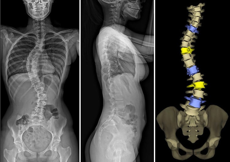
EOS images of a patient with scoliosis: 2D anteroposterior (frontal) and lateral (outer side) X-ray views, and 3D anteroposterior rendering of the spine and pelvis.
On this page:
What is EOS imaging?
EOS imaging is a low-dose, weight-bearing X-ray technology. It can simultaneously take full-body, frontal and lateral (side view) images of the skeletal system of a patient in a standing or sitting position, using significantly less radiation than traditional X-rays or CT scans.
Using EOS, two dimensional (2D) and three-dimensional (3D) orthopedic images can be produced to assist doctors with the diagnosis and treatment of medical conditions of the spine, hips and knees.
What is EOS imaging used for?
EOS imaging is used for anatomical assessment of the entire musculoskeletal system and is invaluable for evaluating the following conditions:
- scoliosis
- kyphosis
- limb length discrepancy
- balance and posture complications
- hip dysplasia
- bowleg and knock knee conditions
Schedule EOS imaging at HSS. (A prescription is required.)
EOS can also support orthopedic surgeons with their presurgical planning, because it records and displays the patient’s anatomical structures in their true, size and volume, lengths and angles. This allows surgeons to perform highly precise presurgical planning and postsurgical assessment for hip replacement and knee replacement surgeries.
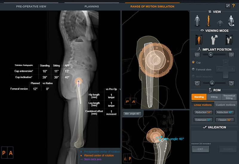
EOS preoperative imaging of a patient with 3D renderings and measurement of a hip replacement implant.
How does EOS imaging work?
During an EOS exam, the patient stands or sits in an upright position inside a special scanning cabin. Two very narrow X-ray beams – one vertical, one horizontal – scan entire body to create 2D and 3D images of the spine and joints.
Unlike traditional X-ray imaging, where the patient may have to be repositioned to get views from different angles, these two simultaneous scans provide all the imaging necessary. Capturing frontal and lateral (side-view), full-body images takes less than twenty seconds. If a full-body image is not necessary (such as for a knee condition), the EOS system can be set to scan a particular region of the patient’s anatomy.
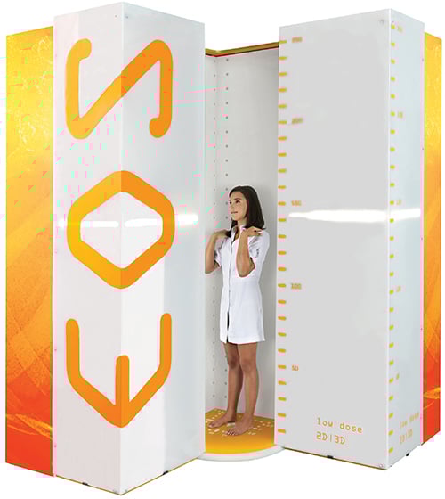
An adolescent patient standing in the EOS low-dose imaging cabin.
How does EOS reduce radiation exposure?
- EOS uses a Nobel Prize-winning detector design capable of capturing more photons (X-rays) than a conventional X-ray detector. Because fewer photons are required to produce the diagnostic image, fewer X-rays are needed to create a high-quality image.
- Very thin slots collimate the X-ray beam. This means that the particles of its rays are accurately parallel and travel in a narrow path, which exposes the patient to only a minimal number of X-rays.
- Technical parameters of the acquisition are adjusted by highly skilled radiology technologists for the size and age of the patient to maintain the lowest dose possible.
How much radiation does an EOS scan emit?
A typical EOS scan at HSS has a radiation dose equivalent to about one-third that of the dose of a conventional X-ray of the same body parts.
EOS is a pediatric-friendly scan. For some children and teens who require frequent imaging to monitor progress of a chronic skeletal or musculoskeletal condition (such as scoliosis), multiple doses of radiation from standard X-rays may unnecessarily increase their life-long radiation exposure. EOS can effectively replace standard X-rays in these instances and thereby reduce any risks associated with radiation exposure over their lifetime.
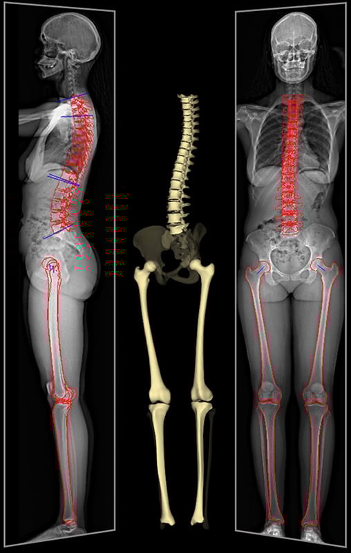
EOS full-body imaging of a patient showing 2D X-ray images and 3D rendering of the spine, hips and leg bones.
Can EOS replace CT scans?
In some cases, an EOS scan can be used instead of a CT scan. For example, precision measurements of a patient’s leg length can be measured from an EOS scan in place of a CT scanogram, which is traditionally used to measure limb length discrepancy.
Advancements in clinical use cases for EOS continue to be investigated, and there may be additional CT scan alternatives soon.
Which HSS locations have EOS imaging?
EOS imaging is available at HSS in New York City, White Plains, NY, and Paramus, NJ, in these locations:
- HSS Radiology – 3rd floor, main hospital building
- HSS Radiology – 2nd floor, The Pavilion
- Lerner Children’s Pavilion, 5th floor, main hospital building
- HSS Westchester, serving Westchester and lower Fairfield County, CT
- HSS Paramus – Midland Ave, serving northern New Jersey and Rockland County, NY
Key takeaways
- EOS imaging produces full-body, frontal and side-view images of the skeleton while patients stand or sit upright.
- The technology uses significantly less radiation than traditional X-rays or CT scans, making it especially beneficial for pediatric patients who need frequent imaging.
- EOS creates both 2D and 3D images that allow precise measurement of bones, joints, and alignment.
- Common uses include evaluating scoliosis, kyphosis, limb length discrepancy, hip dysplasi, and posture-related issues.
- Surgeons use EOS scans to plan and assess procedures like hip and knee replacements with high accuracy.
- EOS exams are quick (under 20 seconds for a full-body scan) and can be targeted to specific body regions if needed.
- In some cases, EOS can replace CT scans, such as for measuring limb length discrepancies.