CT Scan (Computed Tomography)
To diagnose certain injuries and conditions, a doctor may order CT scan (also known as a "CT exam" and commonly, if incorrectly, called a "CAT scan") to obtain two-dimensional images of bones, organs or other body tissues. Learn more about CT scans and how they are used.
What is a CT scan?
A CT scan (or computed tomography examination) is an advanced, noninvasive radiological exam that provides a 360-degree X-ray view of a selected area of the body. It uses high-speed computers and a series of thin X-ray beams to obtain multiple digital images of the area of the body scanned.
What is a CT scan used for?
A CT scan is used to help doctors diagnose abnormalities of bone and soft tissues by producing multiple, layered images of the area of the body that is scanned. These layers of two-dimensional (2D) images are arranged similarly to slices of a loaf of bread.
Taken from multiple angles, these images can provide a three-dimensional (3D) view of the area scanned. A CT scan is a highly sensitive method of accurately viewing internal anatomy and detecting extremely small areas of damage (lesions) caused by injury or disease.
Schedule a CT scan at HSS. (A prescription is required.)
Benefits of a CT scan vs. a standard X-ray
- CT scans have shown to be a cost-effective imaging tool for a wide range of clinical problems.
- CT scanning is painless, noninvasive and accurate.
- New state-of-the art CT scanners produce superior exams using a fraction of time and radiation exposure. At HSS, we practice ALARA* protocols for all our patients, including in children. New technologies make faster scanning possible. For children this means shorter imaging times and less time required to hold still in order to produce clear, crisp images. Also, shorter scan times will make it easier for children and adults to hold their breath during critical parts of the exam. Scanning helps to reduce the need for sedation in children.
- A major advantage of CT is its ability to image bone, soft tissue and blood vessels all at the same time.
- Compared to MRI, CT is less sensitive to patient movement and can be performed if you have an implanted medical device of any kind.
- CT imaging provides real-time imaging, making it a good tool for guiding minimally invasive procedures such as needle biopsies and needle aspirations. Sometimes ultrasound is substituted for CT as a method of image guidance for procedures, especially in children.
- A diagnosis determined by CT scanning may eliminate the need for exploratory surgery and surgical biopsy.
Why did my doctor order a CT scan?
A CT scan may be requested in order to assess bones, soft tissues (such as cartilage, muscle, and tendons) and/or joints for damage, lesions, fractures or other abnormalities. This is especially true when other types of examinations, such as an X-ray, MRI and/or physical exam have not produced a conclusive diagnosis.
CT provides highly detailed information about bony structures, joints, soft tissue and soft tissue calcifications. At HSS, the most advanced technology is used to diagnose abnormalities of the spine, the bones, joints, in addition to abnormalities of the abdomen, pelvis and head.
Special protocols are used for detailed imaging of the spine, the pelvis, the hips, the shoulders, the bones and joints of the extremities. CT is used to gain additional detailed information after myelograms, discograms, and after selected types of arthrograms. CT is also used to guide procedures such as biopsies, aspirations of fluid collections, to guide facet joint injections and nerve root blocks.
Who performs and interprets the CT scan?
The CT scan is ordered by your referring physician and is interpreted by a radiologist. A radiologist is a physician with dedicated training in the safe utilization of imaging equipment and cross-sectional image interpretation. The radiologist will protocol (prescribe) the specific examination parameters. A specially trained technologist will operate the CT scanner according to the prescribed protocol. The radiologist will supervise and confirm that the examination is performed accurately, interpret the study, and provide a written report to your physician. All radiologists at HSS are certified by the American Board of Radiology and have experience and/or fellowship training in orthopedic conditions.
What is special about CT scans at Hospital for Special Surgery (HSS)?
HSS uses the most advanced CT scanning technologies available. In addition, our radiologists who analyze and interpret CT scans are uniquely qualified for sub-specialization of diagnosing orthopedic and musculoskeletal conditions.
Benefits of having a CT scan at HSS
- The most highly skilled radiology technologists perform the prescribed musculoskeletal scans.
- The radiologists who protocol and interpret your CT scan are sub-specialized in conditions of the musculoskeletal system and orthopedic conditions.
- HSS CT scans are specialized for fine osseous (bony) and soft tissue detail scanning.
- HSS has multiple, customized orthopedic CT scan protocols designed specifically for the individual orthopedic subspecialties of our different surgical divisions and centers.
- Same day service is available, and accommodation of patients into the appointment schedule on an as-needed basis.
- Immediate reading results are available. (These are also known as "stat" results or "wet reads", since prior to digitalization, X-ray films were reviewed immediately after being removed from developing solutions. Today, the imaging is 100% digital.)
- Stealth scanning for virtual spine surgery can be performed upon request.
- 3D images are created upon physician request to enhance anatomical visualization of fractures/dislocations. This technology can be used to optimize visualization of complex fractures and or dislocations.
- Specialized studies may be performed to evaluate limb rotational abnormalities (anteversion and retroversion studies) and limb length discrepancy.
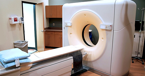
How is a CT scan performed?
An X-ray tube and a series of detectors are contained in a doughnut-shaped machine. You will lie down on a platform that will move you slowly through this doughnut. Some CT scan examinations will require an injection of an iodinated contrast agent (dye) into your vein. If contrast is needed, an intravenous (IV) line will be placed for the injection. Patients undergoing CT-guided biopsies, joint injections and/or aspirations, facet and/or nerve root injections may have local anesthesia and needle placement, as required, for performing these specific procedures.
What should I do to prepare for the examination?
If your CT scan will be performed as part of another procedure (surgery, biopsy, etc.), follow the doctor's instructions for that procedure. If the CT scan is to include an injection of a contrast agent, it is advisable that you do not eat or drink for a several hours before the procedure. If you have any allergies, especially allergies to medications, contrast agent, local anesthesia, Betadine soap, or latex, be sure to inform your physician at the time of scheduling of the procedure, and also inform the CT technologist and the radiologist before the start of the procedure.
If you are pregnant or think you may be pregnant, be sure to inform your physician, the technologist, and the radiologist prior to the procedure. Most procedures/examinations using X-rays will not be performed on pregnant women unless the benefits of the procedure/examination outweigh the risks of radiation exposure to the fetus.
What are the risks of a CT scan?
CT scans expose patients to radiation, but they are within the limits determined to be acceptable (safe) by the National Radiation Safety Commission. If intravenous (IV) contrast dye is required, there is a risk of allergic reaction, as well as minor bleeding or soreness at the contrast injection site.
Reduction of radiation exposure
Although many X-ray image slices may be obtained on a portion of your body, each is done so with a very thin X-ray beam in order to minimize exposure and scatter the radiation. The X-ray exposure you may receive will be within the safety limits put forth by the National Radiation Commission. HSS radiology equipment is monitored daily by highly trained technologists and a radiation physicist. When appropriate, radiation-sensitive of the body will be shielded during the scan.
Contrast risks
- Contrast reactions: Some patients may have an allergic reaction to the contrast agent. In most people who do, these reactions are relatively minor, such as itching, hives, nausea or vomiting. Less frequently, more serious allergic responses can include anaphylactoid reaction, hypotension (low blood pressure), shock and, in rare cases, death.
- Allergies: If you have a history of prior reaction to a contrast agent, be sure to inform your physician at the time of scheduling of the CT scan. Also, at the time of the examination, be sure to inform the technologist and the radiologist performing the exam prior to the injection of any contrast. Individuals with a history of prior severe contrast reaction will either have the exam performed without contrast or in certain cases will have it performed with contrast after a course of premedication with steroids and antihistamine. The premedication is started the day before the exam.
- Bleeding/soreness: Bleeding may occur at the puncture site for the contrast dye IV line, and some patients may experience soreness at the site, similar to having a vaccination or other type of injection. There may also be soreness, swelling and/or bruising for up to a few days at the site of any site of needle placement for any related biopsy, joint injection and/or aspiration, facet injection or nerve root block.
What are the alternatives to a CT scan?
Possible alternatives may include routine X-rays, ultrasound images or MRIs. For certain spine conditions, low-radiation EOS imaging may also be an option. Your physician will order a CT scan only when the specific information a CT offers will be useful toward making a diagnosis, planning treatment, or following the progress of treatment.
What can I expect after getting a CT scan?
If intravenous contrast dye was used, you may have soreness at the site of IV line for up to a few hours. If a biopsy, joint injection, and/or aspiration, or nerve root block was performed, soreness at the site of needle placement may last up to a few days or a hematoma with swelling and a black and blue appearance can develop at the site of needle placement. If you had a therapeutic injection you will receive a discharge instruction form.
What happens with the results?
A radiologist (medical doctor with specialized training in medical imaging) will interpret your CT scan write a report of the same. This report will become a part of your medical record and a copy will be sent to the physician who ordered the CT scan. If an abnormality of urgent nature is discovered on your scan, the radiologist will inform your physician immediately. Copies of the report can be obtained through your referring physician's office. Your physician can call the file room at 212.606.1015 and a copy of the report can be faxed or mailed free of charge to their office. The images obtained are the property of the institution as are biopsy slides or blood samples. Copies of the images can also be obtained by contacting the file room. There is a charge for obtaining film copies and mailing them to your physician. To protect our patients private health information, all patients requesting images and/or reports are required to first complete a Request for Release of Information and fax it to 212.774.2327.
Will other tests be ordered?
Depending on your symptoms, condition or results of the CT scan, your physician may order additional types of imaging studies in order to further evaluate your condition.
Articles related to computed tomography
Read articles about how CT scans are used to help diagnose different conditions.
In the news
Updated: 7/26/2022
References
- *ALARA is an acronym for "as low as (is) reasonably achievable." This means making every reasonable effort to maintain exposures to ionizing radiation as far below the dose limits as practical as defined under federal guidelines.