Bone Lesion
Medically reviewed by Taylor J. Reif, MD
Growth abnormalities in bone tissue known as bone lesions may be benign or malignant.
- What is a bone lesion?
- What is the difference between a benign and a malignant bone lesion?
- What is a bone tumor?
- Who gets benign bone lesions and who gets malignant lesions?
- What causes bone cancer?
- How are benign and malignant lesions diagnosed?
- How are benign lesions treated?
What is a bone lesion?
A bone lesion is any process that replaces normal healthy bone with abnormal bone or tissue. The abnormality will fall along a spectrum ranging from tissue that closely resembles normal bone and which are no cause for alarm, to that which is very distinct from bone and worthy of further investigation to determine a diagnosis and guide treatment. A major categorization of a lesion is whether it is benign or malignant.
What is the difference between a benign and a malignant bone lesion?
Benign lesions form in a bone and can grow locally but do not spread to other organs to cause harm.
Malignant lesions, more commonly referred to as cancer, are lesions which may form and develop in the bone but have the capacity to spread to other areas of the body and continue to grow. For bone cancers, this most commonly occurs to the lungs, where growth can lead to difficulty breathing and ultimately prove fatal.
In some cases, cancers originating in other parts of the body can migrate (metastasize) into bone tissue. This is known as a metastatic lesion.
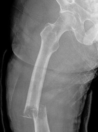
Example of a metastatic lesion in the femur (thighbone), leading to a pathologic fracture
What is a bone tumor?
A bone lesion is considered a bone tumor if the abnormal area has cells that divide and multiply at higher-than-normal rates to create a mass in the bone. The term "tumor" does not indicate whether an abnormal growth is malignant (cancerous) or benign, as both benign and malignant lesions can form tumors in the bone. Types of cancerous bone tumors include:
- osteosarcoma (also known as primary bone cancer) in which a metastatic lesion originates in the bone
- metastatic bone disease (also known as secondary bone cancer) in which cancers originating in other parts of the body invade bone tissues
Who gets benign bone lesions and who gets malignant lesions?
Benign bone lesions can be due to genetic causes, growth disturbances, or changes in the behavior of a small group of cells. Growth disturbances are abnormal changes in a child’s growth pattern, and can be caused by skeletal dysplasias, metabolic disorders and various organ conditions.
We often don’t know why a lesion forms in a specific area of bone in a specific person, but over time physicians have developed an understanding of how the lesions behave regardless of why they formed. Benign lesions usually develop during growth and development, and so they are often diagnosed in younger patients. However, lesions that have not caused symptoms may be first identified well into adulthood.
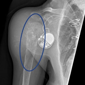
Anteroposterior (front-to-back) X-ray view of a chondrosarcoma (malignant bone tumor) in right proximal humerus (upper arm bone). The patient’s pacemaker for an unrelated heart condition is also visible.
Malignant bone lesions, or bone cancer, occurs when a cell in the bone is able to multiply without restraint and evade the body’s defenses to destroy this abnormal tissue. Our understanding of bone cancer continues to progress, but there is no simple way to predict who will develop bone cancer or when it will develop. Certain rare conditions like Paget’s disease or Maffucci’s syndrome place a person at higher risk to develop bone cancer, but most develop spontaneously.
What causes bone cancer?
Bone cancer is caused by mutations in a person’s DNA. These mutations can be genetic, spontaneous or induced by environmental factors. Bone cancer affects all age groups, but certain subtypes, like osteosarcoma and Ewing sarcoma are more common in children and adolescents, while chondrosarcoma is more common in adults.
How are benign and malignant lesions diagnosed?
Lesions in the bone are usually identified on radiographic images – chiefly X-rays – but also on CT and MRI scans. For those that are possibly cancerous, a biopsy is conducted to identify it.
Imaging is often helpful in determining a diagnosis, and it can sometimes make a particular diagnosis nearly certain. However, many lesions look quite similar and no diagnosis can be made immediately.
For lesions that look concerning for cancer or are causing damage to the bone, a biopsy is performed to make a specific diagnosis, which guides appropriate treatment.
For lesions that do not appear cancerous or destructive, multiple images over time can be obtained and compared for changes, and biopsies are often unnecessary.
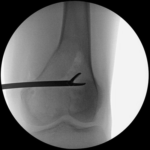
X-ray example of a biopsy being performed on a lesion in the distal femur (lower thighbone). In this image, a bone forcep is removing a piece of the lesion to be sent for laboratory testing.
How are benign lesions treated?
Many benign lesions are stable in the bone and require no treatment. Some benign lesions or tumors can be locally aggressive and require surgical treatment to eradicate the process and prevent further damage.
How are malignant lesions treated?
Malignant lesions always require treatment. Malignant lesions are usually treated with surgery to remove the tumor, but they may also require other forms of treatment, such as chemotherapy or radiation therapy.