Osteosarcoma (Bone Cancer)
Medically reviewed by Taylor J. Reif, MD
What is osteosarcoma?
Osteosarcoma is a form of cancer that originates from within a bone. Unlike many conditions labeled "bone cancer" – which are actually cancers that originate in other organs (such as the breast, prostate or lung) and then spread to bone tissue (known as secondary bone cancer) – osteosarcoma is a malignancy of the bone itself (also known as primary bone cancer).
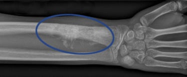
X-ray image of an osteosarcoma in the distal radius (the upper portion of the larger of the two forearm bones).
The cancer cells in osteosarcoma (also sometimes called osteogenic sarcoma) produce an immature and nonfunctional form of bone osteoid that invades and replaces normal bone tissue. The presence of this abnormal bone matrix aids in making the diagnosis both on radiographic imaging and in the pathologic examination of the cells in the lab.
Who gets osteosarcoma?
Classic osteosarcoma is diagnosed in children, adolescents and teenagers. However, older individuals can develop a form of the disease as well. When older people are diagnosed with osteosarcoma, it is often associated with other medical conditions, such as Paget’s disease, or instances in which the area of the body has been treated previously with radiation therapy.
What are the symptoms of osteosarcoma?
The main symptom of osteosarcoma is typically pain near a joint (most commonly the knee), which is not relieved by rest or pain medications. The pain usually increases with time and is aggravated by impact activity (such as running) or, sometimes, merely by putting weight on the extremity.
How is osteosarcoma diagnosed?
An X-ray is the first step in making a diagnosis. Once there is suspicion for an osteosarcoma, it is recommended that the patient be referred to a physician who specializes in orthopedic oncology for additional tests, including a biopsy.
The orthopedic oncologist will order MRI imaging of the suspicious area of the bone. These advanced images will guide the physician in performing a biopsy of the bone lesion. The biopsy is a surgical procedure that obtains a sample or core of the bone which can be further examined under a microscope by a pathologist.
When a diagnosis for osteosarcoma is made, further imaging studies will be performed to determine whether the cancer has spread elsewhere in the body.
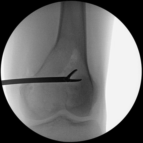
X-ray example of a biopsy being performed on a lesion in the distal femur (lower thighbone). In this image, a bone forcep is removing a piece of the lesion to be sent for laboratory testing.
How is osteosarcoma treated?
The treatment of osteosarcoma is conducted by a team of doctors primary lead by an oncologist and an orthopedic surgeon. The oncologist directs the chemotherapy treatment of the disease which is indicated in most forms of osteosarcoma. The orthopedic surgeon performs the removal of the tumor and the reconstruction of the limb. There are many options for surgical treatment, so it is essential that both the patient and family have significant input to determine the best approach. New techniques to reconstruct the affected bone with the patient’s own live bone are possible in many circumstances, even during chemotherapy.
After the initial chemotherapy and surgical procedures, the patient is followed over time to monitor for disease recurrence. Although any diagnosis of cancer is life-altering for a patient and their family, osteosarcoma is treatable and many patients are cured of the disease.