Progressive Hip Pain, Stiffness, and Neurologic Symptoms in a 26-Year-Old Man with Skeletal Dysplasia
From Grand Rounds from HSS: Management of Complex Cases | Volume 14, Issue 2
Case Report
A 26-year-old man with skeletal dysplasia presented with progressive bilateral hip pain, stiffness and weakness, and abnormal gait. He had right-sided genu valgum, a limb-length discrepancy, and limited hip range of motion. His height was 5′2″, and a marked thoracic kyphosis, mild scoliosis, and increased lumbar lordosis were noted. Staged bilateral total hip arthroplasty (THA) was recommended, after which the limb-length discrepancy would be addressed. He was referred to the Kathryn O. and Alan C. Greenberg Center for Skeletal Dysplasias (CSD) and for high-risk anesthesia consultation.
He first began experiencing low back and hip pain at 8 years old, and over several years his height moved from the 10th percentile down to the 3rd percentile. He received leuprolide to delay puberty and growth hormone to increase height. Childhood X-rays showed vertebral and epiphyseal changes consistent with spondyloepiphyseal dysplasia (SED), but results of genetic testing in childhood for X-linked SED tarda and urinalysis for metabolic syndromes were normal.
At the CSD, the patient underwent genetic testing that detected compound heterozygous GLB1 gene variants, consistent with a diagnosis of GM1 gangliosidosis/Morquio syndrome type B, subsequently confirmed by reduced β-galactosidase enzyme activity (3.19 nmol/hr/mg protein). This diagnosis matched the patient’s signs and symptoms: childhood growth delay, progressive pain and stiffness, spondyloepiphyseal dysplasia, dysplastic vertebrae with anterior beaking and wedging, scoliosis, early-onset osteoarthritis, and neurologic conditions (dystonia, gait disturbances, speech difficulty) [1].
The patient’s surgical history included bilateral femoral derotation osteotomies and removal of hardware, after which he was diagnosed with restrictive lung disease. At HSS, radiographs showed bilateral hip dysplasia with severe osteoarthritis and mild superolateral subluxation of the left femoral head (Figure 1). Dedicated spine views showed thoracolumbar scoliosis, anterior wedging of T11 with focal kyphosis, and severe lumbar disc height loss (Figure 2). There was no cervical spine instability on flexion and extension views (Figure 3).
Figure 1. X-ray shows bilateral hip dysplasia with severe osteoarthritis.
Figure 2: X-ray shows thoracolumbar scoliosis, focal kyphosis at T11, and lumbar disc height loss.
Figure 3: X-ray shows no cervical spine instability.
He was referred to a neurologist because of a persistent pulling sensation on the left-side of the face and left-hand stiffness; other neurologic symptoms included a tendency to startle (with or without stimulus), slurred speech, dizziness with intermittent blurred vision, and underlying dysplasia. Neuraxis magnetic resonance imaging (MRI) detected canal stenosis of the thoracic and lumbar spine, but neurologic exam detected no evidence of myelopathy or lumbosacral radiculopathy.
To limit the risk of nerve impingement on the day of left THA, the patient self-positioned in a lateral decubitus position of comfort that was replicated during the procedure. Given the spinal canal stenosis, a general anesthetic was used.
A posterolateral approach to the left hip was used, as was robotic-assisted navigation to allow placement of the acetabular component with maximum bone coverage. The patient had a flatback deformity but normal spinal mobility, and so the cup was placed in 42° of inclination and 22° of anteversion. The leg was short due to joint degeneration and femoral head subluxation, and a 10-to-15-mm limb lengthening was planned. The prior rotational osteotomy resulted in 3° of femoral retroversion; the stem was templated with 15° of anteversion vs the native neck (Figure 4). This led to a downsizing of the uncemented femoral stem to prevent impingement. A size-3 high-offset stem with + 0 mm head increased the leg length by 12 mm and restored offset (Figure 5).
Figure 4: The prior rotational osteotomy resulted in 3° of femoral retroversion; the stem was templated with 15° of anteversion vs the native neck.
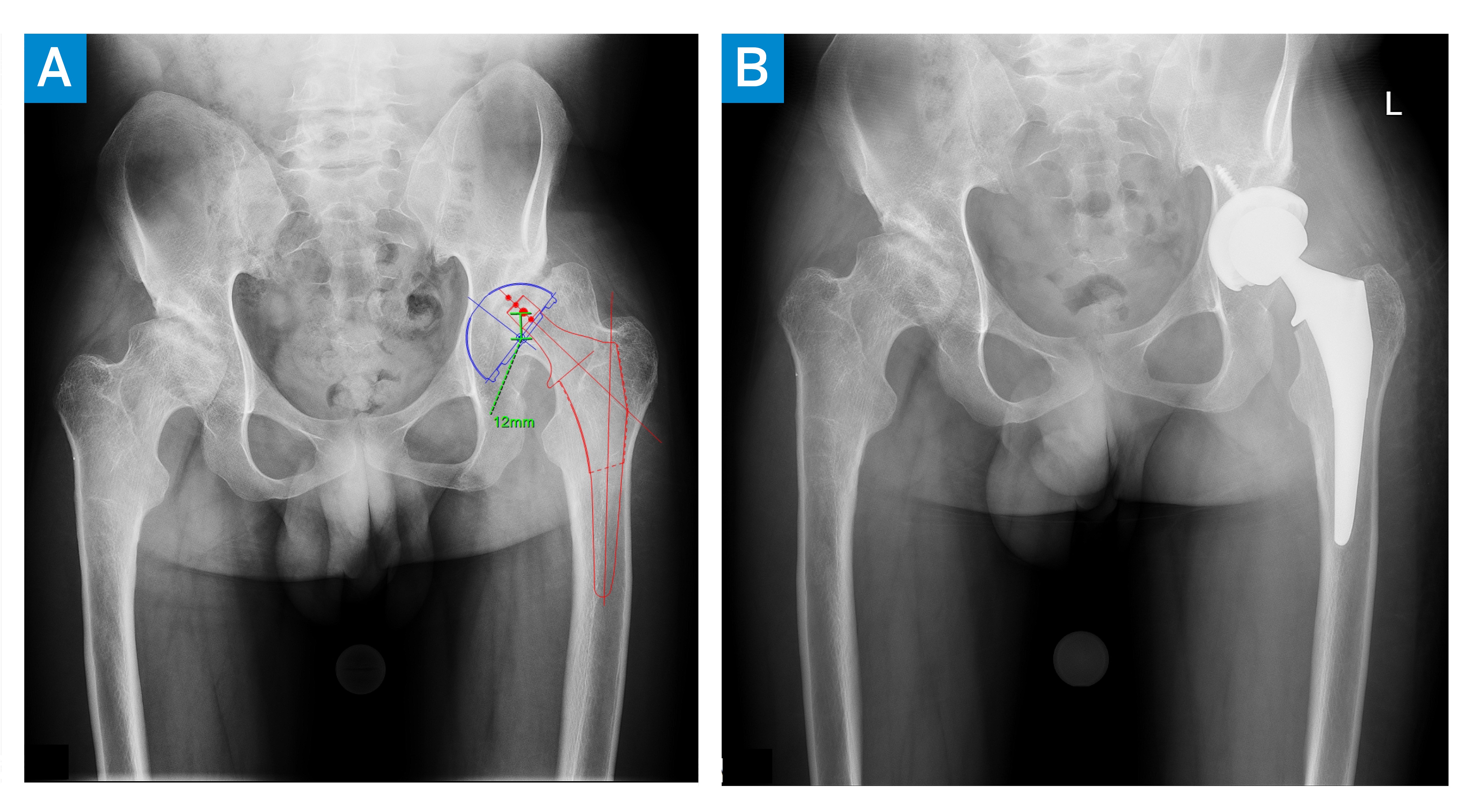
Figure 5: Leg length was restored by 12 mm.
The patient was placed on posterior hip precautions for 6 weeks postoperatively and during physical therapy noted a dramatic improvement in gait. The left hip flexion contracture resolved, improving pelvic position and torso posture, and he could use his abductors to stabilize the pelvis and increase stride length. THA of the right hip was planned, after which leg length discrepancy and valgus deformity of the right leg could be addressed.
Discussion
This adult patient’s clinical history and X-ray findings seemed to match a diagnosis of a lysosomal storage disorder, a group of rare genetic disorders characterized by the progressive dysostosis multiplex, a skeletal dysplasia affecting the joints, the spine, and the ligaments. Some of this patient’s neurologic features overlapped with both Morquio B syndrome and the adult-onset GM1 gangliosidosis [1, 2].
As this case demonstrates, accurate diagnosis may not be achieved until an individual reaches adulthood [3], and many patients with Morquio syndrome require THA by the second decade of life [4, 5]. The risk for spinal cord compression must be evaluated preoperatively [5], and extra-skeletal risks for airway management and cardiopulmonary complications require thorough preoperative clearances, ideally with a multidisciplinary team with experience in managing these complicated disorders [6].
Authors


References
- Brunetti-Pierri N, Scaglia F. GM1 gangliosidosis: review of clinical, molecular, and therapeutic aspects. Mol Genet Metab. 2008;94(4):391-396. doi: 10.1016/j.ymgme.2008.04.012.
- Stockler-Ipsiroglu S, Yazdanpanah N, Yazdanpanah M, et al. Morquio-like dysostosis multiplex presenting with neuronopathic features is a distinct GLB1-related phenotype. JIMD Rep. 2021;60(1):23-31. doi: 10.1002/jmd2.12211.
- Prat C, Lemaire O, Bret J, Zabraniecki L, Fournié B. Morquio syndrome: diagnosis in an adult. Joint Bone Spine. 2008;75(4):495-8. doi: 10.1016/j.jbspin.2007.07.021.
- Abumansour IS, Yuskiv N, Paschke E, Stockler-Ipsiroglu S. Morquio-B disease: Clinical and genetic characteristics of a distinct GLB1-related dysostosis multiplex. JIMD Rep. 2019;51(1):30-44. doi: 10.1002/jmd2.12065.
- Yuskiv N, Higaki K, Stockler-Ipsiroglu S. Morquio B disease. Disease characteristics and treatment options of a distinct GLB1-related dysostosis multiplex. Int J Mol Sci. 2020;21(23):9121. doi: 10.3390/ijms21239121.
- Theroux MC, Nerker T, Ditro C, Mackenzie WG. Anesthetic care and perioperative complications of children with Morquio syndrome. Paediatr Anaesth. 2012;22(9):901-7. doi: 10.1111/j.1460-9592.2012.03904.x.