Lupus Blood Test Results Explained
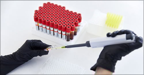
Patients with suspected or confirmed systemic lupus erythematosuss ("lupus" or SLE) undergo laboratory tests (blood tests) for multiple reasons. Physicians and other health care professionals test patients periodically and use the information derived from the tests in various ways.
Uses for laboratory tests
- To diagnose: Lupus symptoms often mimic those of other diseases and vice versa. Physicians use medical history, physical examination and laboratory findings to confirm the diagnosis.
- To determine prognosis: Physicians want to understand how a patient’s disease will progress. Laboratory tests are used to establish a baseline at the time of diagnosis and to predict whether lupus is likely to improve or worsen.
- To monitor: Laboratory tests help physicians assess the severity of the disease, the efficacy of treatment, and medication-related side effects.
- To guide therapy: Laboratory tests help physicians make treatment recommendations and adjust for changing symptoms.
Laboratory tests for autoantibodies in lupus
Antinuclear antibody (ANA)
Antinuclear antibody (ANA) autoantibodies, or antibodies produced by the immune system that attack the body’s own cells, are a hallmark of lupus. ANA is usually measured as 0 to 4+ or as a titer (the number of times a blood sample can be diluted and still be positive). ANA of 0, 1+ or 2+, or at titers less than 1:80 (diluted 80 times) usually do not indicate a significant problem. ANA titers at higher levels more likely indicate the presence of autoimmune disease. Many laboratories also measure pattern or the way the test looks when viewed through a microscope. Different ANA patterns may correspond with different characteristics of lupus.
ANA is a screening test, since almost all patients with lupus have a strongly positive test. However, a positive ANA test result does not by itself confirm a diagnosis for lupus. About 10% of people who do not have an autoimmune disease, as well as many who have other autoimmune diseases (such as thyroid disease) also have positive ANA tests, but usually less strongly positive. Once a person has a positive result, their ANA generally remains positive, so future ANA tests need not be repeated.
Anti-double-stranded DNA (dsDNA)
The anti-double-stranded DNA (dsDNA) antibody is named for its ability to bind to the normal DNA in patients’ cells. At high titers, this antibody is almost exclusively specific to people who have lupus. Low titers can occur in other rheumatic diseases, like rheumatoid arthritis and primary antiphospholipid syndrome. Sometimes, family members of people who have lupus may also test positive themselves, even though they do not have lupus. About 80% of patients with active, untreated lupus have a positive anti-dsDNA test. Monitoring anti-dsDNA is important, since levels may vary along with symptoms of disease, high titers indicating active disease, low titers quiescent disease. (However, anti-DNA antibody levels remain high in some patients with lupus who are well and do not need treatment.) Laboratories vary in how they report the test. Some do so as a range of 0 to 4+ (0, 1+, and 2+ are low titers; 3+ and 4+ are high), others as numbers. To know what is considered high you have to know the range used by the laboratory that conducted your test results.
A diagnosis of lupus is based on a combination of symptoms, physical examination abnormalities (for example, a rash or swollen joints), and laboratory tests. Not all patients with lupus have the anti-dsDNA antibody. Patients who have lupus but do not have anti-dsDNA often have a related antibody, anti-Sm.
Anti-Smith (anti-Sm)
Anti-Smith (anti-Sm), named for the first person known to have this antibody, is the antibody seen in most patients with lupus who do not have anti-dsDNA. Some patients have both anti-dsDNA and anti-Sm. Anti-Sm antibody binds to a protein that is attached to DNA. Unlike anti-dsDNA, the Sm antibody does not change in titer during a lupus flare or in response to treatment so need not be monitored.
Anti-RNP
Anti-RNP (anti-U1 ribonucleoprotein) is a non-specific antibody that occurs in many patients with lupus and other rheumatic diseases. When present in high titer − again, check how the laboratory reports its values to interpret the test − and in the absence of other autoantibodies, anti-RNP suggests a specific “lupus-like” disease, called mixed connective tissue disease (MCTD), characterized by symptoms of lupus, scleroderma, and dermatomyositis, that include:
- puffy hands or other scleroderma-like features
- Raynaud's phenomenon (fingers and toes become numb and pale in response to cold temperatures or stress)
- pulmonary hypertension (high blood pressure that affects the arteries in the lungs and the right side of the heart)
- rashes
- muscle inflammation and weakness
Patients with MCTD, unlike those with SLE, have low risk for developing kidney disease.
Anti-Ro/SSA and anti-La/SSB
Anti-Ro/SSA and anti-La/SSB antibodies identify other molecules in the cell nucleus. Two different groups of researchers discovered these antibodies almost simultaneously. One named them anti-Ro and anti-La for the first letters of the names of the patients in whom they were found; the other group named the antibodies SSA and SSB for Sjogren’s Syndrome A and B, since they are characteristic of this illness. Most doctors use both names.
For technical reasons based on the size of the antigens (molecules to which the autoantibodies react), anti-Ro/SSA is subdivided into 60kd (kilodalton, the size of the molecule) and 52kd components, and La/SSB is 48kd. Distinguishing among them is useful in some special circumstances.
Patients with these antibodies:
- May develop Sjogren’s Syndrome, an autoimmune disorder characterized by dry eyes, dry mouth and arthritis.
- Are more likely to suffer from sun-sensitive rashes.
- May have infants who develop neonatal lupus.
- The mild form of neonatal lupus − which affects up to 20% of infants of pregnant women with lupus who have high titer anti-Ro/SSA and/or anti-La/SSB antibodies − is characterized by rash, sometimes with abnormal blood counts or liver function tests, lasts a few weeks and usually needs no treatment.
- Less than 2% of infants of antibody-positive women develop a serious heart condition. The heart condition occurs mostly in infants of women who are strongly positive for all three 60, 52, and 48kd antibodies. For this reason, fetuses of pregnant women with this antibody profile are closely monitored during pregnancy with noninvasive tests.
Antiphospholipid antibodies
Antiphospholipid antibodies (aPL) occur in about one-third of lupus patients. About 10% of lupus patients have antiphospholipid syndrome, which is characterized by the presence of aPL along with recurring blood clots, pregnancy complications, and other clinical features. Laboratory tests that identify aPL are:
- lupus anticoagulant (LAC), a test for blood clotting (sometimes referred to by the specific test done, such as aPTT or dRVVT)
- anticardiolipin (aCL), which has three subcomponents: lgG, lgA, lgM
- anti-Beta-2-glycoprotein-I (aβ2GPI), which has the same three subcomponents
LAC
The LAC is the most important of these antibodies because it confers the greatest risk for developing clinical complications. Of the subcomponents of aCL and aβ2GPI, IgG is associated with the greatest risk. As with other autoantibodies, strongly positive tests are much more important than are weakly positive ones. LAC may be reported as a ratio (1:3 is low positive, 1:5 or 1:6 is strong positive) or as seconds needed to form a clot, compared to normal done that day (if the normal is 28 seconds, 35 seconds and higher would be unequivocally positive and 60 seconds very strongly positive).
aCL and aβ2GPI
For aCL and aβ2GPI, using the standard international units, normal is usually 16 to 20, equivocal 21 to 40, positive 41 to 80, and high positive (greater than 80). Because a number of infections and injuries can cause temporarily positive antibodies that are not an indication of disease, a diagnosis of antiphospholipid syndrome usually requires that the antibody be present for at least 12 weeks (as indicated by a repeat positive test checked at least 12 weeks after the first).
If aPL are found in a patient who has no blood clots and is not pregnant, usually no treatment is warranted, though individual circumstances may suggest use of aspirin or hydroxychloroquine and sometimes other medications if risk of blood clot is high, for instance before a long airplane flight or surgery. If a patient has a blood clot or other signs of antiphospholipid syndrome, anticoagulants (blood-thinning medications) may be advised, potentially throughout the patient’s life.
Autoantibody panels
Commercial laboratories sell panel tests, combining several of the above tests and sometimes other tests that are not routinely done, that these labs say give better information and more conveniently than do standard tests. As of this writing, however, there is no proof that these tests are beneficial, because false positives are common.
Additional tests used to monitor lupus and general health
The tests below help to monitor disease activity and guide treatment. If the results are abnormal, further medical evaluation may be warranted to determine whether the abnormality is due to lupus, medications used to treat it, or some unrelated health problem.
Complement level: C3, C4, and CH50
The complement system is a series of proteins that assemble in domino fashion to destroy bacteria and viruses invading the body. The signal that initiates this domino cascade, or “activates” complement, is when an antibody meets an antigen (the bacterium or virus, when this occurs in the context of a healthy immune system protecting the body from harm). Because lupus autoantibodies initiate the same signal and activate complement despite the absence of an identified bacterium or virus, the measurement of complement proteins can be used to monitor lupus activity. It usually suffices to measure only two of the complement proteins, called C3 and C4. CH50, or total complement, is a blood test that measures all the complement proteins at once and may give additional information. CH50 is a much more complicated test, however, and is not routinely done.
Low C3 and C4 levels (below 60 for C3 and below 15 for C4 in the usual American measure), occur in active lupus, especially when the kidneys are affected or there is immune breakdown of blood cells (autoimmune hemolytic anemia, AIHA). Other manifestations of lupus, such as brain disease, do not cause low complement levels.
Low complement levels are not specific to lupus. They may be seen in other immune-system-mediated illnesses and in severe infections. About 10% of patients with lupus are born with abnormal complement components, especially C4, so their tests are always abnormal. Interpreted in context, C3 and C4 levels may reflect the activity of the disease. In most patients, recovery from lupus flare is indicated by the return of complement levels to normal. In an apparently well patient who usually has a normal complement level, a sudden decrease may signal an impending flare and the need for close physician follow-up. But a decrease in complement level does not itself warrant treatment, if no symptoms are present.
Complete blood count (CBC)
CBC provides information about the red blood cell (RBC), white blood cell (WBC), and platelet counts, and health of RBCs, all of which may be abnormal in lupus and may need treatment. Common issues are:
- Anemia (low RBC, as measured by hematocrit and hemoglobin). A normal hematocrit is 35% to 40%, hemoglobin 11.5 to 15.0. In patients with lupus, anemia can be caused by active disease, AIHA, bleeding, drug toxicity and, sometimes, by genetic abnormalities like thalassemia or sickle cell disease.
- Leukopenia (low WBC). A normal WBC count is 4,500 to 10,000. Almost all untreated patients with lupus have leukopenia − it can be a clue to the diagnosis. WBCs protect against infection. Abnormally low WBC can be due to lupus, drug toxicity, and certain infections. A mildly low count is not concerning, but if it is below 2,000, the patient is at risk for infection.
- Thrombocytopenia (low platelets). A normal count is 150,000 to 300,000. Thrombocytopenia is also common in lupus and can be caused by the disease, medication toxicity, and other illnesses. Counts between 50,000 and 100,000 serve as an alert to the physician to monitor the patient closely. When platelet counts are below 30,000, the patient is at risk for hemorrhage and likely requires treatment.
Erythrocyte sedimentation rate
Erythrocyte sedimentation rate (ESR, also sometimes called simply "sedimentation rate" or "sed rate") is an indirect indicator of inflammation. The test measures the rate (in millimeters per hour) at which red blood cells (erythrocytes) settle to the bottom of a test tube of blood. If inflammation is present, these cells can stick together and settle more quickly than individual cells. A high ESR, if there are no other reasons for it to be high (such as infection), suggests that lupus is active. A low rate is reassuring. The test is simple but not indicative of any one specific disease, and it is subject to inaccuracy. Normal values may range from 0mm/hr to 20 mm/hr − or 0mm/hr to30 mm/hr, depending on the laboratory – and the interpretation also varies based on age, sex, and other factors.
C-Reactive protein (CRP)
CRP, like the ESR, is an indirect indication of inflammation, but is more specific in the detection of disease activity, since it is not affected by as many variables. Levels may be high in obese persons and in infection. A normal value is less than one milligram per deciliter (<1.0 mg/dL).
Confusingly, cardiologists use a measure called high-sensitivity CRP (hsCRP) to predict risk of heart disease. The hsCRP test is the same as a CRP test, but done differently. It is roughly 10 times more sensitive than is the standard CRP. A result flagged as “abnormal” may not be concerning in a patient known to have SLE or another autoimmune condition.
Complete metabolic panel (CMP) and other common metabolic tests
These laboratory tests look at liver, kidney, endocrine, and other functions:
- Glucose level is a measure for diabetes.
- Sodium (Na), potassium (K), and chloride (Cl), creatinine (Cr) and blood urea nitrogen (BUN) levels measure kidney function.
- Aspartate transaminase (AST), alanine transaminase (ALT), alkaline phosphatase (alkP), bilirubin measure liver function.
- Lactic dehydrogenase (LDH) measures liver function and may be high in forms of illness in which tissue damage occurs.
- Creatine phosphokinase (CPK), or creatine kinase (CK), may be high when there is damage to muscle tissue.
A given laboratory value may have different implications depending on the patient’s presentation. For example, an elevated AST might suggest muscle inflammation, and an elevated LDH and bilirubin might suggest AIHA. As with all laboratory tests, accurate interpretation requires guidance from a physician or other healthcare professional with a complete understanding of the clinical context.
Urinalysis with microscopy
Because lupus patients are prone to kidney disease, urine tests are ordered on a regular basis. Urine tests are evaluated for:
- pH − To determine if the urine is acidic (normal) or alkaline (suggests infection or problems in the way the kidney functions).
- Protein − The level should be 0 or trace. If protein is present, it may indicate kidney dysfunction.
- Protein/creatinine ratio − This is a test to quantify the protein in the urine. Higher ratios are more suggestive of kidney dysfunction. Sometimes a 24-hour urine collection is done to confirm the results.
- WBC − Only a low number (less than five) are normally present. A high WBC count in urine may indicate a urinary infection.
- Blood − More than a very small number of RBC or a positive test for hemoglobin may indicate bladder or kidney disease or kidney stones (unless a woman has her menstrual period when the sample is collected).
- Casts − Uncommon, but certain types may suggest more severe kidney disease.
- Bacteria – May indicate infection if the specimen was properly collected.
In summary
It is important to note that:
- No two patients with lupus are the same.
- Healthcare professionals will take into consideration the whole person and context when diagnosing and treating lupus.
- Lab tests can help complete the puzzle of diagnosis, treatment and monitoring of the disease.
- Each lab test provides information about different components of lupus and can help doctors better understand the characteristics of lupus in a given patient.
- Patients should communicate with their doctors and ask about their lab test results in order to understand their disease.
Learn more information about the SLE Workshop, a free support and education group held monthly for people with lupus and their families and friends.
