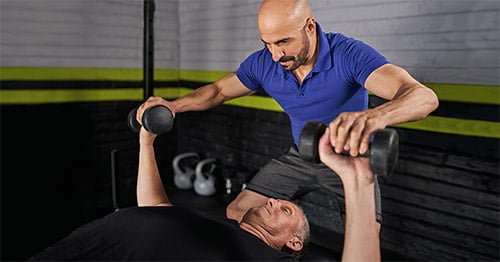
Shoulder Arthroscopy
Medically reviewed by Russell F. Warren, MD
- What is shoulder arthroscopy?
- Anatomy of the shoulder joint
- What shoulder conditions can be treated arthroscopically?
- How long will I be in the hospital?
- What type of anesthesia will I have?
What is shoulder arthroscopy?
Shoulder arthroscopy is a minimally invasive technique that allows orthopedic surgeons to assess – and in some cases, treat – a range of conditions affecting the shoulder joint.
During the procedure, the orthopedic surgeon makes small incisions or portals in the affected joint, and then inserts a tiny camera and fiber optics to light the interior space. Pictures obtained with the camera are then projected onto a screen in the operating suite.
Whenever possible, arthroscopic techniques are preferred to traditional, open shoulder surgery, which involves larger incisions and longer recovery times.
Anatomy of the shoulder joint
While many people think of the shoulder as a single joint, it is actually made up of two joints: the acromioclavicular joint, where the acromion of the shoulder blade and the collarbone (clavicle) meet, and the glenohumeral joint, where the head of the humerus (the upper bone in the arm) meets the glenoid, the cup-like portion of the scapula.
There is also potential space (the subacromial space) between the acromion and rotator cuff tendon. Injuries to the shoulder may occur in either joint or in the soft tissues that support and stabilize it.
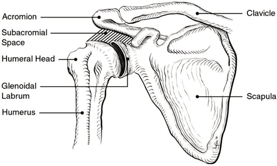
Bones of the shoulder
What shoulder conditions can be treated with arthroscopic surgery?
Conditions in the shoulder that may be diagnosed and treated with the help of arthroscopy include biceps tendon and rotator cuff injuries, shoulder impingement, labral tears, frozen shoulder, arthritis and other problems.
Shoulder conditions that may benefit from arthroscopic surgery
- Acromioclavicular joint (AC joint) injuries, which may require stabilization with ligament reconstruction.
- A torn labrum of the shoulder.
- Shoulder instability, which may be secondary to a torn labrum or a loose joint capsule (capsular laxity). Both can be repaired with sutures and anchors to rebalance the joint.
- Articular cartilage injuries, and, where appropriate, use of articular cartilage regeneration techniques.
- Frozen shoulder, a condition of unknown origin in which the patient develops synovitis and subsequent contracture of the shoulder joint capsule, resulting in a very limited range of motion. In order to restore mobility, the orthopedic surgeon makes small cuts in the tissue to release the contractures.
- Biceps tendon conditions, including:
- Biceps tendon disease causing pain. An arthroscopic tenotomy can be performed to release the tendon.
- A frayed biceps tendon may be treated with debridement (a smoothing of rough surfaces, and removal of loose tissue).
- A rupture of the biceps tendon where it connects the muscle to the shoulder may be treated with biceps tenodesis surgery, which may also involve a debridement of the bicipital groove of the humerus (upper arm bone), followed by suturing of the tendon to the groove.
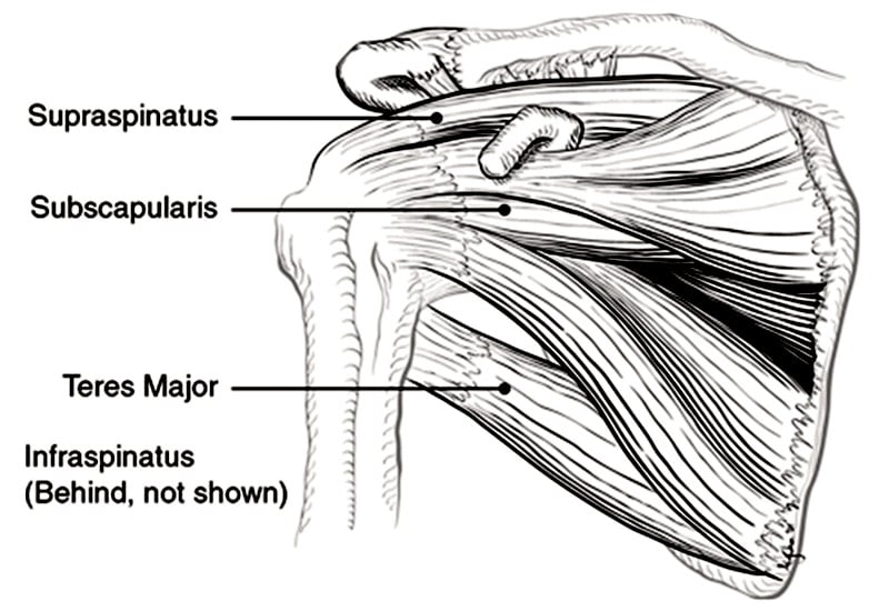
- Rotator cuff conditions, including:
- Tendonitis and tears of the rotator cuff. Tendonitis and some partial tears (depending on depth) can often be treated arthroscopically with debridement or tendon repair. For repair, sutures attached to anchors allow the surgeon to advance the tendon to bone and promote healing.
- Shoulder impingement: a condition in which the rotator cuff tendon becomes inflamed or abraded. If a narrow subacromial space (the gap between the acromion and the glenohumeral joint below it) is damaging the tendon, the acromion may also be thinned arthroscopically to allow more space. A bursectomy may also be needed.
- Calcium deposits in the rotator cuff, which can cause pain and stiffness; excision can provide relief.
- Arthritis of the shoulder: debridement of cartilage and loose tissue bodies in younger patients can provide symptom relief, but only for a variable period of time. If pain returns over time, a shoulder replacement may be the only surgical option. If a person with severe arthritis of the shoulder has also had a prior rotator cuff tear, they may need to have a procedure known as reverse shoulder replacement.
Shoulder arthroscopy may be used for smaller fractures to aid in repair, such as tuberosity fractures and anterior glenoid fractures. Shoulder infections may be debrided and washed out via shoulder arthroscopy.
Diagnostic shoulder arthroscopy
View this animation to learn how shoulder conditions may be diagnosed using arthroscopy.
How long will I be in the hospital after shoulder arthroscopy?
Shoulder arthroscopy is typically ambulatory, which you go home the same day of the procedure.
What type of anesthesia will I have for shoulder arthroscopy?
Arthroscopic shoulder procedures may be performed under either regional anesthesia or general anesthesia, depending on the case. Regional nerve blocks are associated with fewer complications. Arthroscopic shoulder surgeries at HSS are most frequently performed using regional anesthesia and mild sedation. General anesthesia is usually not necessary.
Learn more about shoulder arthroscopy from the content below.
In the news
- Shoulder Pain? Chances Are It's Your Rotator Cuff
- Reverse, anatomic TSA had similar return to sport, satisfaction rates
- Shoulder Replacement Surgery: Signs, Types and Recovery Guide
- David M. Dines, MD, Receives Lifetime Achievement Award from Long Island Business News
- Liposomal Bupivacaine Shows Similar Length of Pain Relief as Standard Bupivacaine With Dexamethasone After Shoulder Surgery
- Anatomic TSA still relevant as use of RSA increases
- Recurrent instability similar with arthroscopic Bankart repair, open bone block procedure
Explore Related Patient Stories
View All Patient Stories
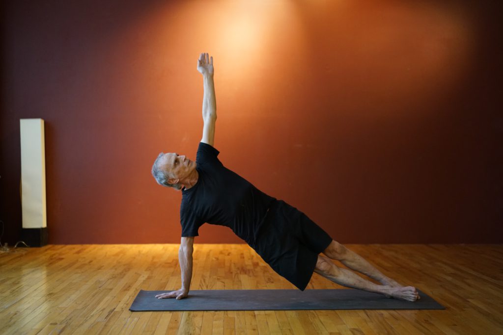
Carl Joshua Smith
New York, NY
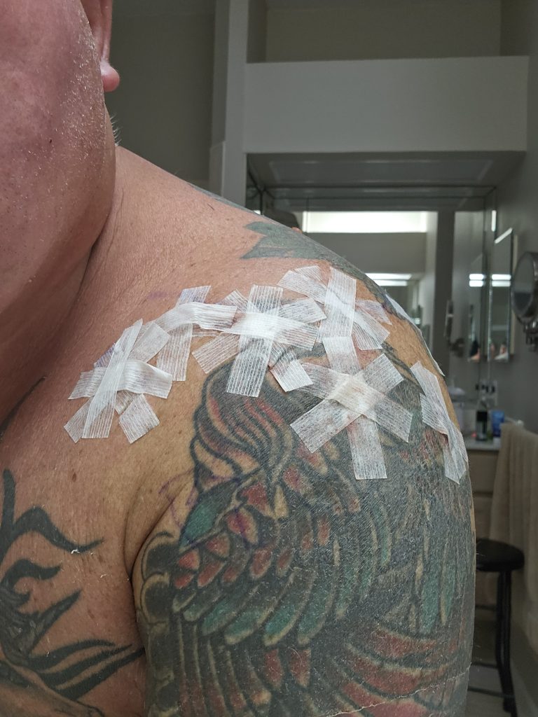
Shoulder Arthroscopy
Salvatore Burriesci
Port St. Lucie, FL
Shoulder Arthroscopy
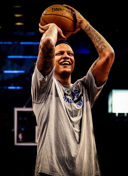
Charlie Villanueva
Dallas, TX
Shoulder Arthroscopy
Frederick Len Butler
Babylon, NY
Shoulder Arthroscopy

Russel Matson
Margaretville, NY
Shoulder Arthroscopy
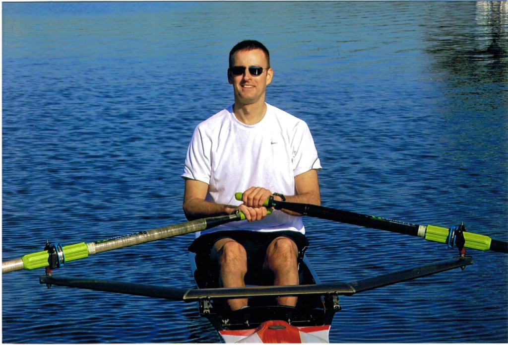
Thomas Lutazi
New York, NY
Shoulder Arthroscopy

Steve Messenger
New York, NY
Shoulder Arthroscopy
Karen Swartz
Middletown, CT
Shoulder Arthroscopy
Richard Mayer
Waltham, MA
Shoulder Arthroscopy

Howard Herman
Brooklyn, NY
Shoulder Arthroscopy
Adam Sokolow
New York, NY
Shoulder Arthroscopy
Kim Thompson
Mason Neck, VA
Shoulder Arthroscopy
Quilvio Cabral
Santo Domingo, Dominican Republic
Shoulder Arthroscopy

Gorm Amand
New York, NY
Shoulder Arthroscopy
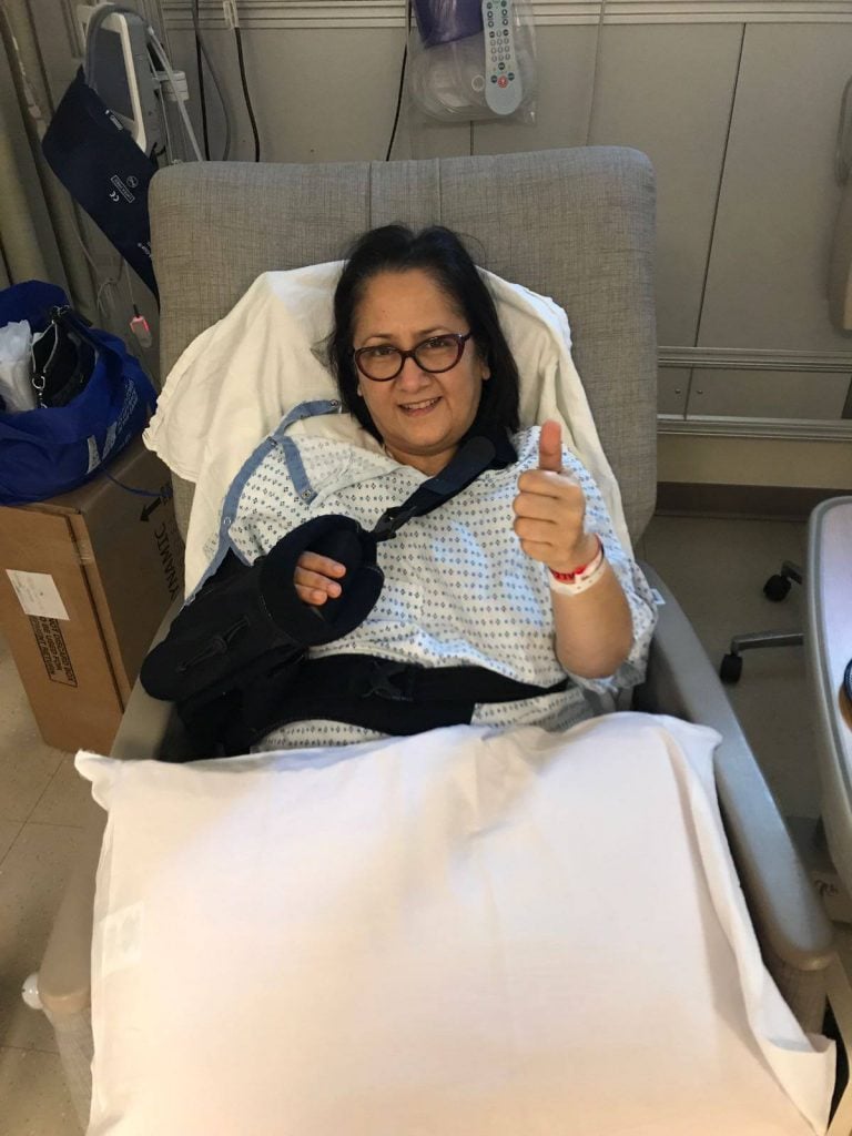
Sumita Bhattacharya
White Plains, NY
Shoulder Arthroscopy
Sumie Okazaki
New York, NY
Shoulder Arthroscopy
Michael Engelmann
New York, NY
Fractures of the Shoulder and Collarbone

Stephen Machnicki
Weston, CT
Shoulder Arthroscopy
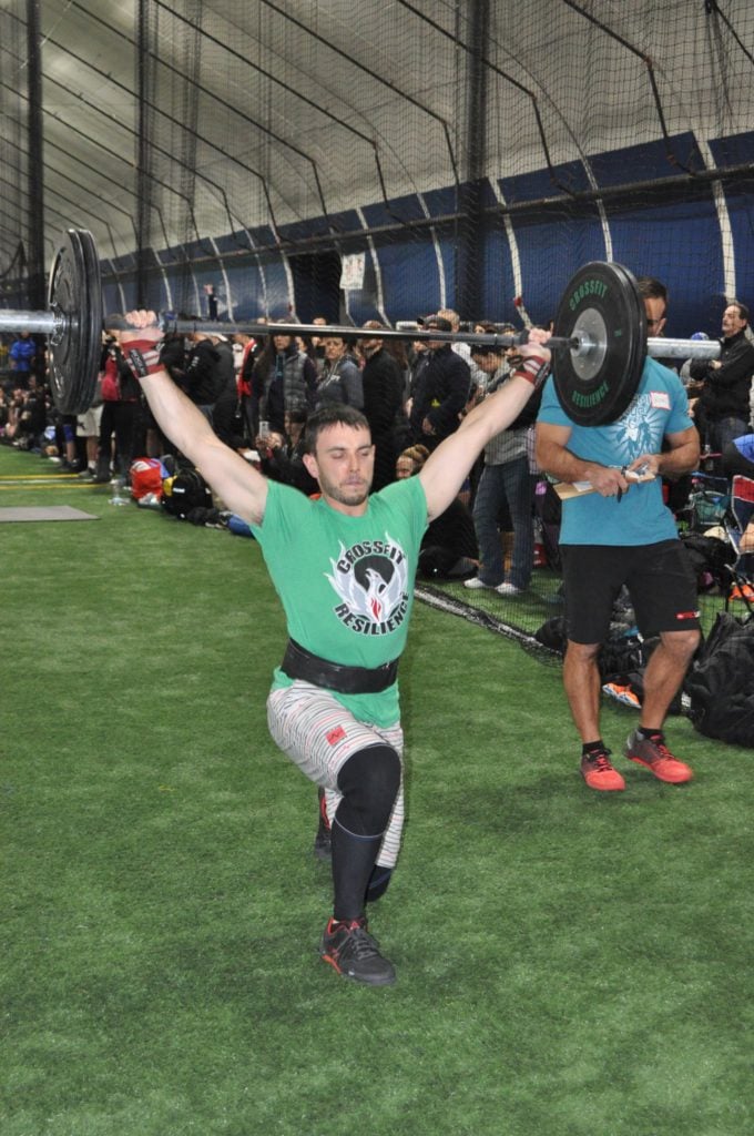
Kristen Tomlinson
Weehawken, NJ
Shoulder Arthroscopy
Greg Vigilante
Waterford, NY
Shoulder Arthroscopy
References
- Apostolakos JM, Wright-Chisem J, Gulotta LV, Taylor SA, Dines JS. Anterior glenohumeral instability: Current review with technical pearls and pitfalls of arthroscopic soft-tissue stabilization. World J Orthop. 2021 Jan 18;12(1):1-13. doi: 10.5312/wjo.v12.i1.1. PMID: 33520677; PMCID: PMC7814310.
- Amirtharaj MJ, Wang D, McGraw MH, Camp CL, Degen RA, Dines DM, Dines JS. Trends in the Surgical Management of Acromioclavicular Joint Arthritis Among Board-Eligible US Orthopaedic Surgeons. Arthroscopy. 2018 Jun;34(6):1799-1805. doi: 10.1016/j.arthro.2018.01.024. Epub 2018 Feb 21. PMID: 29477607.
- Bovonratwet P, Fu MC, Pathak N, Ondeck NT, Bohl DD, Nho SJ, Grauer JN. Surgical Treatment of Septic Shoulders: A Comparison Between Arthrotomy and Arthroscopy. Arthroscopy. 2019 Jul;35(7):1984-1991. doi: 10.1016/j.arthro.2019.02.036. Epub 2019 Jun 10. PMID: 31196694.
- Chen FR, Quan T, Pan S, Manzi JE, Recarey M, Agarwal AR, Nicholson A, Zimmer ZR, Gulotta L, Dines JS. Anesthesia Type and Postoperative Outcomes for Patients Receiving Arthroscopic Rotator Cuff Repairs. HSS J. 2022 Nov;18(4):519-526. doi: 10.1177/15563316221080138. Epub 2022 Mar 3. PMID: 36263279; PMCID: PMC9527545.
- Lamplot JD, Shah SS, Chan JM, Hancock KJ, Gentile J, Rodeo SA, Allen AA, Williams RJ, Altchek DW, Dines DM, Warren RF, Cordasco FA, Gulotta LV, Dines JS. Arthroscopic-Assisted Coracoclavicular Ligament Reconstruction: Clinical Outcomes and Return to Activity at Mean 6-Year Follow-Up. Arthroscopy. 2021 Apr;37(4):1086-1095.e1. doi: 10.1016/j.arthro.2020.11.045. Epub 2020 Dec 2. PMID: 33278535.
- Nicholson AD, Estrada JA, Mathew JI, Finocchiaro A, Pinnamaneni S, Okeke L, Dines DM, Dines JS, Taylor SA, Warren RF, Cordasco FA, Rodeo SA, Gulotta LV. Minimum 15-year follow-up for clinical outcomes of arthroscopic rotator cuff repair. J Shoulder Elbow Surg. 2022 Aug;31(8):1696-1703. doi: 10.1016/j.jse.2022.01.116. Epub 2022 Feb 11. PMID: 35158066.
- Voleti PB, Camp CL, Sinatro AL, Dines JS. Arthroscopic Fixation of Glenoid Rim Fractures After Reduction by Labral Repair. Arthrosc Tech. 2016 Apr 18;5(2):e379-83. doi: 10.1016/j.eats.2016.01.013. PMID: 27462537; PMCID: PMC4947900.