Neck injuries are a particular concern in contact sports such as football, and they require special attention because of the devastating consequences if they are severe. Cervical nerve root and spinal cord injuries are among the most common cervical spine neurologic (nerve) injuries.
The cervical spine refers to the seven cervical vertebras, bones in the neck that connect the back of the skull to the thoracic spine in the upper back. The muscles in the neck support and protect the vertebral bodies, cord, and nerve roots by absorbing forces transmitted to the neck directly, or through the head or trunk.
There are eight cervical nerve roots arising from the cervical spinal cord, which exit the vertebral canal through foramen (circular openings, or "windows", between the vertebras that house the spinal cord). The size or diameter of the vertebral canal and foramen vary in size, depending on the person.
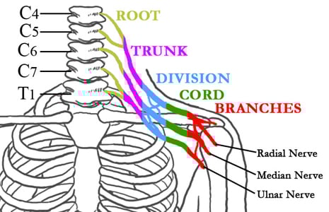
After exiting the canal, the lower cervical nerve roots become interwoven with each other to form the brachial plexus, a series of nerves that supply sensation and motor function to the arms and hands.
Since the diameter of the vertebral canal and foramen differs on an individual basis, a small spinal canal or foramen can put the spinal cord or nerve root at increased risk for injury.
A common neck injury in football involves compression or stretching of a nerve root or the brachial plexus; referred to as “burners” or “stingers”, they make up approximately ten percent of all cervical spine neurologic injuries, and two thirds of all college football players experience at least one of these injuries. These injuries occur from either a pinching (compression) of the nerve on the same side the neck is flexed/extended or a stretching of the nerve on the opposite side of where the neck is flexed/extended.
Those affected may experience sharp, burning pain that may radiate into the shoulder and down the arm to the hand. The symptoms only occur on one side and may include weakness and paresthesia (numbness or tingling) in the involved extremity for several seconds to several minutes. Raising the arm (deltoid), flexing the elbow (bicep), and rotating the arm outwards (using the external rotator muscles of the rotator cuff) may be restricted as a result of motor weakness.
Physical exam should include a thorough sensory exam; a complete motor exam including the deltoid, biceps, and rotator cuff; and a Spurling’s provocative test. A Spurling’s test attempts to reproduce the symptoms of a burner by passive extension and lateral flexion (sideways movement) of the head and neck.
Electrodiagnostic testing or an MR neurogram may be used to identify which nerves are affected. X-rays and an MRI are indicated when there is persistent neurologic deficit or recurrent symptoms, to rule out an occult (hidden) fracture or a herniated disc.
These injuries are more likely to occur in patients with poorly developed neck musculature. In older athletes, nerve root compression is more likely to occur in conjunction with a herniated cervical disc.
Conservative treatment consists of rest, possibly a neck brace, and over-the-counter pain relievers or anti-inflammatory medications. With persistent or recurrent symptoms, it is very important to have a thorough evaluation by an orthopaedic or neurology specialist. Very rarely is surgery indicated to relieve persistent pressure on a nerve, except when there is evidence of small foramen. In this setting, one option is to surgically enlarge these openings to give more space for the nerves.
Much more serious neck injury can result in quadriplegia or quadriparesis. Quadriparesis describes an episode of numbness or tingling, with possible motor change ranging from weakness (quadriparesis) to paralysis (quadriplegia) of all four extremities (arms and legs). These episodes most commonly last from ten minutes to forty-eight hours. There is usually no fracture or dislocation, but the event can be associated with a small (stenotic) spinal canal. With fracture or dislocation of the cervical spine and severe spinal cord injury, quadriplegia may persist and surgery may be indicated.
Quadriplegia or quadriparesis are caused by an acute episode of forced hyperextension, hyperflexion, or excessive loading of the cervical spine. It only occurs in about one out of 10,000 football players every season and is very highly associated with “spear tackling” or other head-first falls. The neck is usually flexed at least thirty degrees, and in this position the cervical spine loses its normal curve, causing the neck musculature to lose its ability to dissipate force.
After this injury, the athlete should not be permitted to return to play until radiographs and an MRI are obtained to prove there is no spinal cord swelling (edema) or instability (ligamentous injury). If these studies are normal, the athlete can return to play when there is painless range of motion of the neck and a return of full strength.
Although there is a fifty percent chance of the injury occurring again after return to play, there is no good data to suggest any long term disability. If the athlete has had multiple episodes or the symptoms last longer than thirty-six hours, they should not be allowed to return to play.
Radiographic proof of a small spinal canal or an episode associated with herniated disk or degenerative changes in the spine, are relative contraindications to return to play. There is some evidence to suggest that people with small spinal canals who experience an episode of transient quadriplegia could be at risk for permanent neurologic problems.
There may be some benefit from giving high dose steroids to the patient at the time of spinal cord injury, but the evidence is not definitive. All of these injuries should be treated with full cervical spine precautions until any fractures, dislocations, or cervical spine instability can be ruled out.
In the setting of football, the helmet and shoulder pads are left in place until cervical spine instability has been ruled out. The facemask may be removed if access is needed to maintain the airway. If the airway appears to be compromised, cardiopulmonary resuscitative efforts should be carried out, but the neck must be stabilized as well. The patient should then be “log rolled” onto a back board and transported to the hospital.
Persistent symptoms with an associated small spinal canal may be treated with a cervical decompression and fusion. However, the extent and specific nature of these injuries may be quite varied; each requiring detailed evaluation and treatment options directed specifically to the injury at hand.
Updated: 10/9/2009
Patrick Birmingham, MD
2008 Sports Medicine Fellow
Hospital for Special Surgery
Or call
+1.877.606.1555