Cartilage Repair and Regeneration
Medically reviewed by Riley J. Williams III, MD
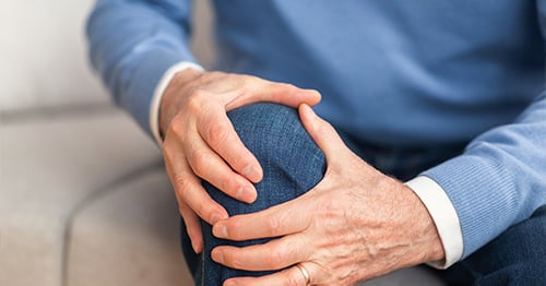
Articular cartilage can be damaged by a traumatic injury, progressive wear or systemic disease. Loss or erosion of articular cartilage causes pain, weakness and dysfunction. If left untreated, cartilage damage can progress and lead to symptomatic arthritis.
Cartilage has a poor capacity to repair itself. As a result, surgery may be necessary for individuals who present with joint (knee, ankle, hip, elbow) dysfunction associated with a painful cartilage or chondral lesion. Successful cartilage repair surgery reduces pain and restores function. There are numerous cartilage repair treatment options available, and the suitability of each of these approaches depends on each patient’s unique clinical problem. Physicians in the HSS Institute for Cartilage Repair are experts in the treatment of damaged articular cartilage. Their clinical approaches are data-driven and, consequently, our physicians note high clinical success rates in their respective patient populations.
What is cartilage repair/regeneration?
In its simplest terms, cartilage repair (regeneration) surgery results in the formation of robust cartilage-like tissue around the joint where the cartilage has worn away or been damaged. Cartilage regeneration typically requires some sort of surgical intervention. Synthetic adjuncts are often needed to successfully regenerate articular cartilage. Adults don’t have the natural capability to grow new articular cartilage from scratch. This ability is only possible in a fetus growing inside the womb. This fact makes cartilage repair surgery challenging.
How is damage to cartilage diagnosed?
Our physicians will take a full medical history, perform a physical exam, and utilize imaging studies (X-rays, MRIs, CT scans) to determine the location and extent of the articular cartilage damage. Advanced MRI technology allows surgeons to design a personalized treatment plan prior to surgery. Continual MRI readings taken after surgery allow for an objective assessment of the performed surgery and the application of a proper rehabilitation plan that, together, facilitate a complete recovery.
Can cartilage heal on its own?
No. Articular cartilage is the smooth cushion that lines the ends of bones where they meet at joints such as the knee, shoulder and ankle. Intact articular cartilage allows bones to move against one another without friction in healthy joints. Cartilage contains no nerves and does not have a full blood supply. As such, cartilage does not have the capacity to heal on its own.
Can damaged cartilage be repaired?
Repair of one’s damaged existing cartilage cannot usually be performed. Yet, there are treatment options in which damaged articular cartilage may be replaced or reconstructed using cell-based or tissue-based strategies.
For example, one commonly performed procedure (mosaicplasty, osteochondral autograft transplant) works by taking small cylinders of cartilage from one area of a patient’s joint (typically a non-weightbearing area) to reconstruct and fill a cartilage defect in a weightbearing area. Another commonly performed procedure uses donated cartilage tissue (osteochondral allograft) to reconstruct large joint cartilage lesions. In yet another method, small arthroscopically harvested cartilage samples can be used to grow a customized cartilage “patch” that can be used to fill a cartilage defect. Surgeons at HSS have been performing these procedures since the 1990s and have been at the forefront of developing all of these described methodologies.
What is cartilage repair surgery?
Cartilage repair surgery consists of regenerating or replacing cartilage, either with tissue from the patient’s own body, someone else’s body, or by generating cartilage repair tissue in a lab.
Who is a good candidate for cartilage repair?
The ideal cartilage repair candidate is someone who suffers from an isolated articular cartilage defect. The patient should have a relatively healthy knee with no generalized cartilage loss (arthritis). The treated knee should be well-aligned and stable (no ligament deficiencies). Cartilage repair can be performed on people of all ages but, typically, patients are under 50 years of age.
What conditions may be treated with cartilage repair?
Cartilage injuries that can be treated by cartilage repair surgery include:
- articular cartilage defects
- chondral defects
- chondral lesions
- osteochondral defects
- osteochondritis dissecans lesions
- osteochondritis dissecans (OCD)
- avascular necrosis (AVN), also known as osteonecrosis
Which body parts can be treated using cartilage repair?
Cartilage repair is most commonly used to treat isolated cartilage defects. Cartilage repair surgery is not performed to treat arthritis. However, cartilage repair surgery may help affected individuals delay or avoid a knee replacement. Cartilage repair may also be used to treat cartilage lesions of the ankle, elbow, shoulder or hip.
What are the types of procedures for cartilage repair?
Due to recent medical advances, there are now multiple treatment options available depending on factors such as size of the cartilage damage and a patient’s end goals.
- Debridement or chondroplasty – The objective of this procedure is to alleviate symptoms associated with the mechanical blocks to motion associated with cartilage lesions. The surgeon will remove the loose fragments of cartilage that are causing joint pain and often send them to the lab to do a cell-based (MACI) procedure later (see below). It may also be beneficial inject an adjunctive treatment, such as bone marrow aspirate concentrate. This is a concentration of the patient’s own bone marrow cells, which aid in healing. Sometimes, a bridging procedure like this is appropriate if the patient is an in-season athlete.
- Microfracture surgery – This arthroscopic bone marrow stimulation procedure involves creating small holes in the base of the cartilage lesion to promote a healing response and create cartilage repair tissue. This is used to treat small areas of cartilage damage and can be effective for short-term treatment of knee cartilage defects while more modern techniques are best for people who seek a durable long-term solution.
- Whole tissue options –
- Osteochondral autograft transplantation surgery (OATS) – Surgeons take articular cartilage from a healthy, non-weight bearing area of the patient’s knee and transplant to the damaged area of articular cartilage. Use of the patient’s own tissue facilitates a very durable repair and excellent clinical outcomes. This method has shown strong results in individuals participating in high-demand activities. This procedure is also known as an autologous osteochondral transfer (AOT).
- Osteochondral allograft transplantation – Surgeons use whole donor tissue specimens to treat large lesions. This method is best for large lesions, and those lesions that also involve large segments of bone (osteochondritis dissecans, avascular necrosis). Osteochondral allograft surgery can also be performed as a salvage procedure for other failed cartilage repair surgeries. Cartilage allograft surgery may not be as durable in the long run in high-demand people
- Matrix-induced autologous chondrocyte implantation (MACI) – Autologous chondrocyte implantation as a long history of clinical success and is one of the most common techniques used today for repairing knee cartilage. In this procedure, surgeons take a small sample of healthy cartilage from the knee in a small surgery. The cells are isolated, grown in a lab, and seeded onto a collagen patch. At surgery, the patch is shaped and glued into cartilage defect. This patch ultimately grows into new, healthy cartilage repair tissue. Research has proven this to be very effective for injuries of the femur (thighbone), patella (kneecap) and tibia (shin bone).
- Particulate juvenile articular cartilage allograft transplantation – This procedure uses small fragments of donor cartilage tissue to facilitate the formation of cartilage repair tissue. Unlike osteochondral allografts, this method has no associated bone with the cartilage. The surgeon transplants small pieces of donated cartilage into the damaged area and secures the transplant with a fibrinous glue. The transplanted cartilage grows quickly into durable cartilage repair tissue. Studies out of HSS have demonstrated the clinical efficacy of this method in high demand individuals.
How long does cartilage repair surgery take?
Most of the described surgical procedures take less than an hour. Surgeries can be performed arthroscopically but may require a small incision to fully execute the procedure. Combining the cartilage repair procedure with other surgeries (ligament reconstruction, osteotomy) may lengthen the procedure accordingly.
How long does it take to recover from cartilage repair surgery?
Most patients use crutches for the first two to three weeks after surgery. Physical therapy usually starts about a week after surgery on an outpatient basis. Most patients can return to normal activities of daily living four to six weeks after surgery. Many patients are cleared for some sports after six months. However, getting back to a high level of fitness or ballistic sports may take longer. Different surgical treatment options have different timetables for a return to high level activities. Osteochondral autograft and allograft patients usually can expect to be cleared at six months. In contrast, the MACI procedure, because this method requires two surgeries spaced six to eight weeks apart, the full recovery time is closer to 12 to 18 months.
Physical therapy is an important part of recovery and should be utilized as appropriate. Postoperative MRIs are used to assess the success of the procedure and show progress through the physical therapy process.
How long before you can return to exercise after cartilage repair?
If you are looking to go back to heavy exercise or athletics, it is important to work with a strength and conditioning coach to help with training.
What is the success rate for cartilage repair?
As a subspecialty, cartilage repair surgery has rapidly evolved since its inception in the late 1990s. Before that time, there were few treatment options available to address this clinical problem. Currently, there are multiple ways to treat cartilage damage. Success rates truly depend on several factors (surgery performed, patient age, body mass index, duration of symptoms etc.). Our surgeons have tracked their clinical outcomes in this area since 1998. The HSS Cartilage Registry tracks the clinical outcomes of over 4,000 patients who have been treated for symptomatic cartilage defects. Our surgeons recommend custom treatment options based on objective data and clinical outcomes.
What are the potential risks of cartilage repair?
There is no more risk involved than typical surgical risk.
Can cartilage be repaired without surgery?
Simply, no. In most cases, surgery is required to repair articular cartilage.
In rare cases, small traumatic cartilage lesions form a repair tissue called fibrocartilage on their own. This typically occurs at the time of injury if there is a significant amount of bleeding and trauma. Fibrocartilage is located between the vertebra of the spine, in the meniscus of the knee, and in joint capsules that surround some joints. Fibrocartilage is inferior to articular cartilage for the purposes of bearing loads in a joint.
What vitamins or supplements help repair cartilage?
Taking vitamins does not help cartilage to repair itself. However, there are supplements that can play a role in controlling and limiting joint inflammation that is associated with cartilage damage. Oral supplements such as glucosamine sulfate or hyaluronic acid can be helpful in relieving symptoms. Neither of these substances have been clinically proven to promote or result in the repair of damaged cartilage. It is our recommendation that it is best to consult with your physician before adding any supplements to your diet.
Learn more about individual cartilage repair surgical procedures and related conditions and treatments in the content below.
References
- Brittberg M, Gomoll AH, Canseco JA, Far J, Lind M, Hui J. Cartilage repair in the degenerative ageing knee. Acta Orthop. 2016 Dec;87(sup363):26-38. doi: 10.1080/17453674.2016.1265877. Epub 2016 Dec 2. PMID: 27910738; PMCID: PMC5389429.
- Crawford DC, DeBerardino TM, Williams RJ 3rd. NeoCart, an autologous cartilage tissue implant, compared with microfracture for treatment of distal femoral cartilage lesions: an FDA phase-II prospective, randomized clinical trial after two years. J Bone Joint Surg Am. 2012 Jun 6;94(11):979-89. doi: 10.2106/JBJS.K.00533. PMID: 22637204.
- Choi YS, Potter HG, Chun TJ. MR imaging of cartilage repair in the knee and ankle. Radiographics. 2008 Jul-Aug;28(4):1043-59. doi: 10.1148/rg.284075111. PMID: 18635628.
- Fernandes TL, Cortez de SantAnna JP, Frisene I, Gazarini JP, Gomes Pinheiro CC, Gomoll AH, Lattermann C, Hernandez AJ, Franco Bueno D. Systematic Review of Human Dental Pulp Stem Cells for Cartilage Regeneration. Tissue Eng Part B Rev. 2020 Feb;26(1):1-12. doi: 10.1089/ten.TEB.2019.0140. Epub 2020 Jan 22. PMID: 31744404.
- Fernandes TL, Gomoll AH, Lattermann C, Hernandez AJ, Bueno DF, Amano MT. Macrophage: A Potential Target on Cartilage Regeneration. Front Immunol. 2020 Feb 11;11:111. doi: 10.3389/fimmu.2020.00111. PMID: 32117263; PMCID: PMC7026000.
- Hede KTC, Gomoll AH, Foldager CB. Demographics in Patients Receiving Matrix-Assisted Chondrocyte Implantation (MACI) in the Ankle. Cartilage. 2021 Dec;13(1_suppl):1331S-1336S. doi: 10.1177/1947603519870854. Epub 2019 Aug 20. PMID: 31431042.
- Hinckel BB, Gomoll AH. Autologous Chondrocytes and Next-Generation Matrix-Based Autologous Chondrocyte Implantation. Clin Sports Med. 2017 Jul;36(3):525-548. doi: 10.1016/j.csm.2017.02.008. PMID: 28577711.
- Hinckel BB, Gomoll AH. Patellofemoral Cartilage Restoration: Indications, Techniques, and Outcomes of Autologous Chondrocytes Implantation, Matrix-Induced Chondrocyte Implantation, and Particulated Juvenile Allograft Cartilage. J Knee Surg. 2018 Mar;31(3):212-226. doi: 10.1055/s-0037-1607294. Epub 2017 Oct 16. PMID: 29036754.
- Hutchinson ID, Moran CJ, Potter HG, Warren RF, Rodeo SA. Restoration of the meniscus: form and function. Am J Sports Med. 2014 Apr;42(4):987-98. doi: 10.1177/0363546513498503. Epub 2013 Aug 12. PMID: 23940202.
- Makris EA, Gomoll AH, Malizos KN, Hu JC, Athanasiou KA. Repair and tissue engineering techniques for articular cartilage. Nat Rev Rheumatol. 2015 Jan;11(1):21-34. doi: 10.1038/nrrheum.2014.157. Epub 2014 Sep 23. PMID: 25247412; PMCID: PMC4629810.
- Krych AJ, Harnly HW, Rodeo SA, Williams RJ 3rd. Activity levels are higher after osteochondral autograft transfer mosaicplasty than after microfracture for articular cartilage defects of the knee: a retrospective comparative study. J Bone Joint Surg Am. 2012 Jun 6;94(11):971-8. doi: 10.2106/JBJS.K.00815. PMID: 22637203.
- Krych AJ, Robertson CM, Williams RJ 3rd; Cartilage Study Group. Return to athletic activity after osteochondral allograft transplantation in the knee. Am J Sports Med. 2012 May;40(5):1053-9. doi: 10.1177/0363546511435780. Epub 2012 Feb 7. PMID: 22316548.
- Mestriner AB, Ackermann J, Gomoll AH. Patellofemoral Cartilage Repair. Curr Rev Musculoskelet Med. 2018 Jun;11(2):188-200. doi: 10.1007/s12178-018-9474-3. PMID: 29777422; PMCID: PMC5970109.
- Moran CJ, Pascual-Garrido C, Chubinskaya S, Potter HG, Warren RF, Cole BJ, Rodeo SA. Restoration of articular cartilage. J Bone Joint Surg Am. 2014 Feb 19;96(4):336-44. doi: 10.2106/JBJS.L.01329. PMID: 24553893.
- Williams, Riley. J., Peterson, L., Cole, B. J., & Humana Press Inc. (2016). Cartilage Repair Strategies.
- Williams RJ 3rd (Riley J. Williams III, MD), Director of the Institute for Cartilage Repair and Attending Orthopedic Surgeon, Hospital for Special Surgery. Video interview, October 14, 2021.