Knee Realignment and Joint Preservation with Proximal Tibial Osteotomy and Cartilage Regeneration
Patient Case History: Richard
Pre-Op
Patient Surgeon: S. Robert Rozbruch
Richard is a 57-year-old man with right knee pain focused on the medial side (inside) of knee. When presented with the option for knee replacement, he was more interested in a joint preservation option.
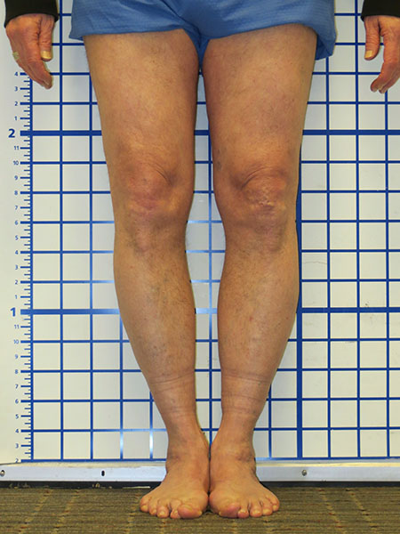
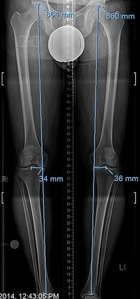
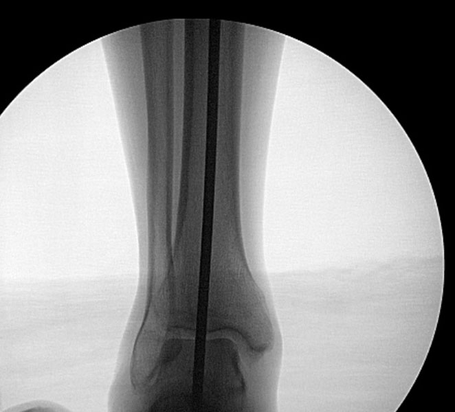
He has a bowleg deformity (Figure 1) which adds excessive stress on the damaged part of the knee. The joint space on the medial side is narrowed from cartilage degeneration (Figure 2).
The long standing x-ray shows that the hip to ankle line passes medial to the inside of the knee indicating overload of the damaged compartment (Figure 3).
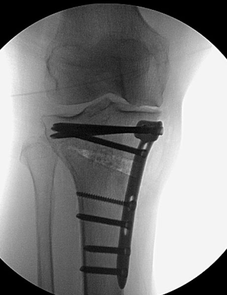
The plan was to correct the bowleg deformity with (medial opening wedge) osteotomy of the proximal tibia and insertion of a titanium plate. This will unload the damaged medial compartment.

Post-Op
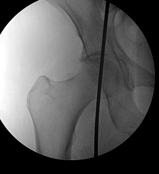
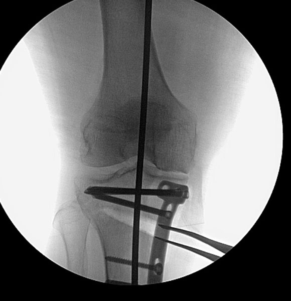
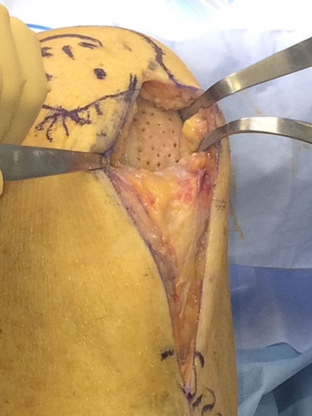
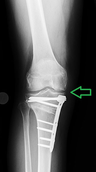
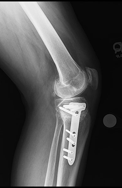
During surgery the optimal re-alignment is confirmed (Figures 1, 2, 3). The opening wedge osteotomy is filled with synthetic graft (Figure 4). Cartilage regeneration of the damaged medial compartment was performed with microfracture technique (Figure 5) combined with insertion of mesenchymal stem cells from iliac crest bone marrow aspiration.
After 6 weeks, Richard can walk without crutches.
Follow-Up
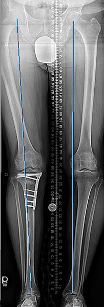
At 3 months, the leg is well aligned (Figures 1, 2), the proximal tibia osteotomy is well healed and the medial joint space and pain are improved (Figure 3, 4). Plans to realign the left leg are underway.

Return to Patient Case Histories