

Severe Sagittal Malalignment in a 43-Year-Old Man with Ankylosing Spondylitis
From Grand Rounds from HSS: Management of Complex Cases | September 2026 Case of the Month
Case Report
A 43-year-old man with human leukocyte antigen (HLA)-B27-positive ankylosing spondylitis (AS) was referred for progressive loss of horizontal gaze. His history was notable for morbid obesity (body mass index, 41.5), obstructive sleep apnea, type 2 diabetes, and a prior small bowel obstruction requiring exploratory laparotomy. He had undergone posterior spinal fusion from T2 to L3 for kyphoscoliosis at an outside hospital in 2007, with extension to C7 in 2008, complicated by a C8 nerve root injury. The management of AS was hindered by intermittent insurance lapses, resulting in prolonged gaps in biologic therapy and periods of medication nonadherence.
He presented with a fixed chin-on-chest deformity with rightward deviation that was not passively correctible. Neurologic examination showed 4/5 strength in the right C8 distribution with no myelopathic signs. He also had a 15° right hip flexion contracture from osteoarthritis.
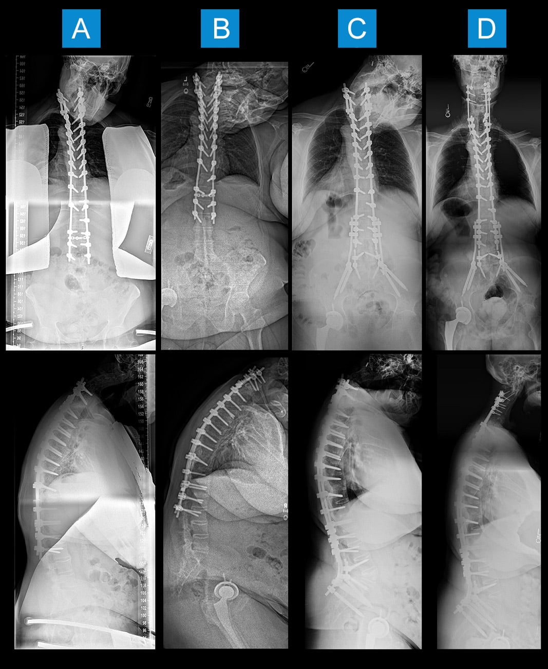
Full-length standing radiographs and computed tomography (CT) demonstrated complete ankylosis from the occiput to the sacroiliac joints, with severe cervicothoracic kyphosis, a T1 slope minus cervical lordosis (TS-CL) of 108°, sagittal vertical axis (SVA) of 160 mm, and a C2-pelvic angle (PA) of 45° (Figure 1A, 1B). The decision was made to proceed with spine surgery first, as the hip flexion contracture was improving with physical therapy and resumed biologic therapy [1]. Preoperative optimization included weight loss, prehabilitation, anesthesiology evaluation for difficult airway requiring awake fiberoptic intubation, and coordination with rheumatology to hold infliximab perioperatively [2].
The first stage consisted of revision posterior spinal fusion from T10 to the pelvis, with an L5 pedicle subtraction osteotomy (PSO). Postoperative radiographs showed correction of SVA from 160 mm to −13 mm, with normalization of regional spinopelvic parameters (L1-PA 2°, T4-PA 3°, C2-PA 17°) (Figure 1C). The patient was discharged to rehabilitation on postoperative day 8.
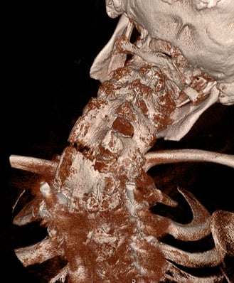
At 8 weeks postoperatively, CT demonstrated a 3-column fracture through C5-6 with distraction of the left-sided C5-6 facet (Figures 2 and 3).
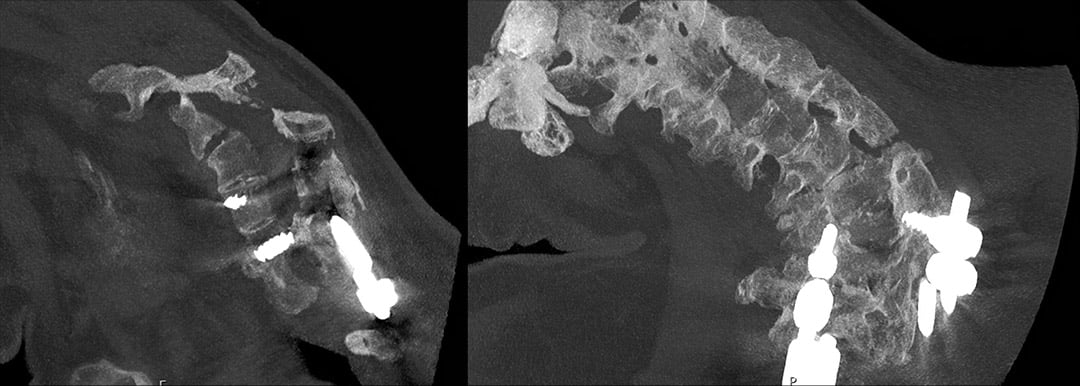
He was admitted and underwent C2-T2 posterior spinal fusion with a C5-6 Smith-Petersen osteotomy for simultaneous fracture stabilization and cervicothoracic deformity correction. Prior C7 and T1 instrumentation was removed, and pedicle screws were placed from C2 to C5. Neuromonitoring remained stable throughout. Postoperative imaging demonstrated correction of TS-CL from 108° to 26°, with further improvement in C2-PA from 17° to 7° (Table 1).
Table 1. Radiographic parameters at 3 timepoints
| Parameter | Preoperative | Post-Stage 1 (L5 PSO) | Post-Stage 2 (Cervical) |
|---|---|---|---|
| Global Alignment | |||
| SVA (mm) | 160 | −13 | 18 |
| C2 Tilt (°) | 17 | 3 | 7 |
| C2-PA (°) | 45 | 17 | 7 |
| Regional Spinopelvic | |||
| L1-PA (°)a | 20 | 2 | 2 |
| T4-PA (°) | 36 | 3 | 3 |
| T4-L1 PA (°) | 16 | 1 | 1 |
| Lumbar Alignment | |||
| L1-4 (°) | 10 K | 10 K | 10 K |
| L4-S1 (°) | 5 | 45 | 45 |
| Regional Cervical | |||
| TS-CL (°) | 108 | 66 | 26 |
| C2-7 Lordosis (°) | 48 K | 48 K | 7 K |
| C2-7 SVA (mm) | 95 | 95 | 45 |
| Pelvic Parameters | |||
| PI (°) | 44 | 44 | 44 |
| PT (°) | 33 | 10 | 14 |
| SS (°) | 11 | 34 | 30 |
|
a Ideal L1-PA = PI/2 − 21° = 44/2 − 21° = 1° for this patient (achieved after Stage 1). Abbreviations: SVA = sagittal vertical axis; C2-PA = C2 pelvic angle; L1-PA = L1 pelvic angle; T4-PA = T4 pelvic angle; T4-L1 PA = T4 to L1 pelvic angle; TS-CL = T1 slope minus cervical lordosis; PI = pelvic incidence; PT = pelvic tilt; SS = sacral slope; K = kyphosis; PSO = pedicle subtraction osteotomy. |
|||
The patient’s postoperative course was complicated by a recurrent small bowel obstruction requiring transfer to an affiliated hospital, where it was managed conservatively. At 3-month follow-up, the patient reported marked improvement in quality of life and was very satisfied with his head position. He denied neck and back pain and had returned to independent activities of daily living.
Discussion
This case highlights several challenges in managing fixed multiplanar sagittal deformity in a patient with an ankylosed spine. We elected to correct the lumbar deformity before the cervical deformity. In a globally ankylosed spine, restoration of lumbar lordosis shifts the trunk’s center of gravity posteriorly, partially compensating for cervical kyphosis and improving global sagittal balance [3]. This staged strategy avoids the morbidity of simultaneous multilevel osteotomies and permits interval reassessment of whether cervical correction remains necessary. Notably, the L5 PSO alone reduced T1 slope from 60° to 18° and SVA from 160 mm to −13 mm, with near-normalization of all regional spinopelvic parameters, demonstrating the profound effect of lumbar correction on global alignment [4].
Ultimately, the presence of a postoperative cervical spine fracture underscores the vulnerability of ankylosed spines to injury from seemingly trivial mechanisms [5]. In AS, the spine behaves as a long bone, and fractures typically propagate through all 3 columns [6]. These injuries are frequently missed and carry high rates of neurologic deterioration, mandating a low threshold for imaging in any AS patient with new or changed symptoms. Here, the fracture provided an opportunity for simultaneous stabilization and deformity correction.
Finally, perioperative management required coordination across rheumatology, anesthesiology, and internal medicine, reflecting the team-based approach essential for complex deformity correction.
AI Use Disclosure
Cursor AI was used to help compile the manuscript using deidentified patient information, generate tables from pre-measured alignment metrics, assist with literature review, and perform formatting and grammar editing. AI was not used to generate clinical images. We reviewed and edited the content and take responsibility for the accuracy of the published article.
Authors


References
- Offierski CM, MacNab I. Hip-spine syndrome. Spine (Phila Pa 1976). 1983;8(3):316-321.
- Goodman SM, Springer BD, Chen AF, et al. 2022 American College of Rheumatology/American Association of Hip and Knee Surgeons Guideline for the Perioperative Management of Antirheumatic Medication in Patients With Rheumatic Diseases Undergoing Elective Total Hip or Total Knee Arthroplasty. J Arthroplasty. 2022;37(9):1676-1683.
- Smith JS, Klineberg E, Schwab F, et al. Change in classification grade by the SRS-Schwab Adult Spinal Deformity Classification predicts impact on health-related quality of life measures. Spine (Phila Pa 1976). 2013;38(19):1663-1671.
- Protopsaltis TS, Scheer JK, Terran JS, et al. How the neck affects the back: changes in regional cervical sagittal alignment correlate to HRQOL improvement in adult thoracolumbar deformity patients at 2-year follow-up. J Neurosurg Spine. 2015;23(2):153-158.
- Shah NG, Keraliya A, Nunez DB, et al. Injuries to the Rigid Spine: What the Spine Surgeon Wants to Know. Radiographics. 2019;39(2):449-466.
- Caron T, Bransford R, Nguyen Q, Agel J, Chapman J, Bellabarba C. Spine fractures in patients with ankylosing spinal disorders. Spine (Phila Pa 1976). 2010;35(11):E458-E464.