

Treating a Child with Severe Neuromuscular Kyphoscoliosis: A 3-Year Journey
From Grand Rounds from HSS: Management of Complex Cases | October 2026 Case of the Month
Case Report
A 4-year-old girl with a history of asthma presented for evaluation of scoliosis. She was first diagnosed at 2 years old at an outside hospital, and bracing had begun at age 3. She had been born via spontaneous vaginal delivery at term and met developmental milestones appropriately. At initial presentation, physical examination was notable only for 1 beat of right ankle clonus. Radiographs showed 78° right thoracic curve, and thoracic kyphosis of 88°, both demonstrating moderate flexibility with traction (Figure 1). Given her age, full spine magnetic resonance imaging (MRI) was obtained; it demonstrated a large syrinx from C2–T10 and a Chiari type I malformation. She underwent an uncomplicated suboccipital craniectomy, C1 and C2 laminectomies, and tonsillopexy with duraplasty.
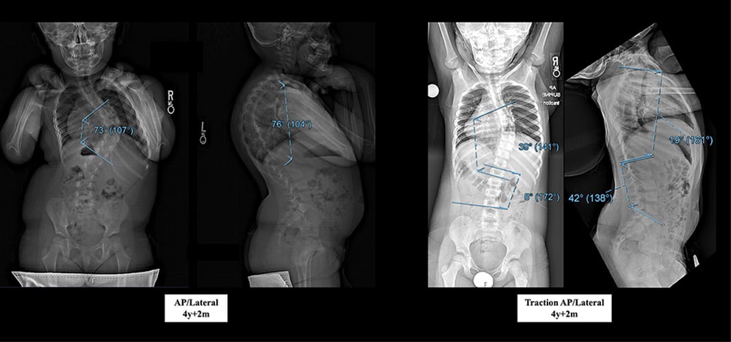
Three months after the neurosurgical procedure, she was placed in a Mehta cast. The patient underwent casting for 1 year, with cast changes every 2 to 4 months (Figure 2).
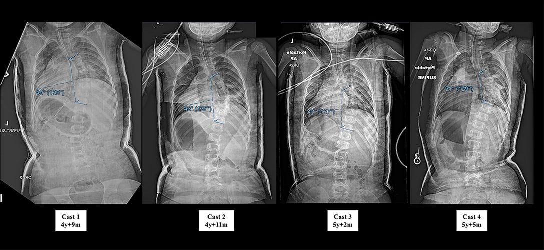
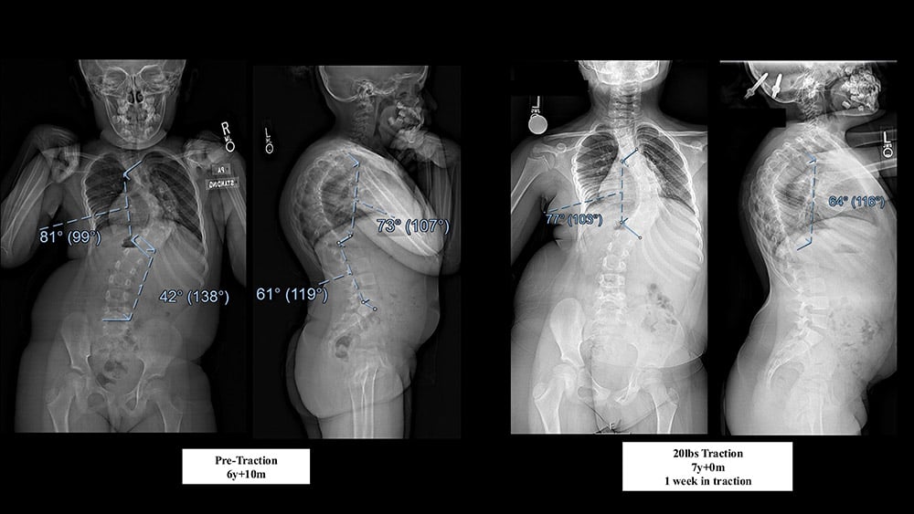
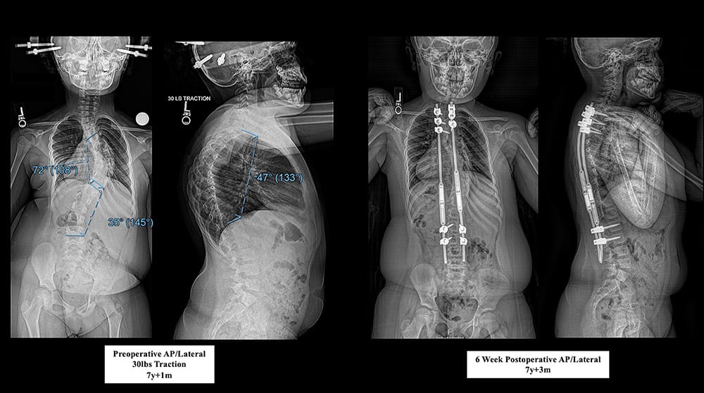
During her fourth cast, she had a severe asthma attack requiring hospitalization, and her casting was discontinued. The casting was successful at stabilizing her curves; she was then transitioned to a custom thoracic-lumbosacral orthosis (TLSO) brace at age 5. She was lost to follow up due to family illness for 8 months, during which time she was not wearing her brace. When she returned at age 6 years, the right thoracic curve had progressed to 83°, with 74° of kyphosis. Given her age and the progression and stiffness of her curve, halo traction and insertion of magnetically controlled growing rods were recommended, which she underwent for 6 weeks shortly after turning 7 (Figure 3). The coronal-plane curve improved minimally, but kyphosis improved significantly to 47°. She underwent insertion of magnetically controlled growing rods from T2–L3, with limited fusion at T2–T4 and L2–L3.
She tolerated the procedure well and was discharged home on postoperative day 3. X-rays 6 weeks later showed intact instrumentation and improvements in thoracic curve to 39° and kyphosis to 35° (Figure 4). At the time of discharge, lengthenings were planned to begin 3 to 4 months postoperatively.
Discussion
Early-onset scoliosis (EOS), defined as scoliosis beginning before 10 years of age, comprises a spectrum of deformities secondary to congenital malformations or neuromuscular conditions, as well as idiopathic causes. Occult central nervous system lesions may exist in up to 22% of cases in patients with curves greater than 20°, despite normal neurological examination [1,2]. Full-spine MRI is recommended for all patients diagnosed with EOS to identify a cause or contributor [3]. In this case, MRI identified a large syrinx and Chiari malformation. Early neurosurgical intervention in patients with a Chiari I malformation associated with syrinx and scoliosis has been shown to improve scoliosis in up to 60% of patients, with curve magnitude over 45° being a negative predictive factor [4].
Progressive EOS before age 5 can impact lung development through limiting both chest wall compliance and the development of alveoli and pulmonary arterioles [1]. Therefore, EOS can result in a restrictive pattern of lung disease due to decreased breathing excursion and ventilation due to lung hypoplasia. If left untreated, severe restrictive disease can lead to thoracic insufficiency syndrome, pulmonary arterial hypertension, and cor pulmonale [1, 5]. In this case, given the patient’s initial diagnosis at 2.5 years of age and asthma history, early intervention was necessary to preserve lung capacity.
The need to preserve pulmonary function is coupled with a need to preserve spinal growth; definitive fusion at an early age results in iatrogenic thoracic insufficiency syndrome [6]. Growing rods (either traditional or magnetically controlled) allow for continued growth in younger children, but both options have been shown to result in decreased overall spinal growth with each lengthening, known as the “law of diminishing returns” [5,7]. This is thought to be caused by progressive stiffening or autofusion around the areas of instrumentation, allowing for only 2 to 3 years of successful growth through growth-friendly constructs prior to fusion [5]. These studies have also demonstrated that revising growing-rod constructs results in continued progressive decline of lengthening capability [5]. Thus, delaying surgical intervention, even one that allows for continued growth, can be useful in delaying final fusion. Mehta demonstrated that serial casting could postpone posterior spinal fusion in EOS patients with large curves (>50°) [8].
This established technique was instrumental in our case, as the patient underwent serial casting for 8 months, before being transitioned to a brace and then undergoing instrumentation [9]. These techniques postponed surgical intervention to age 7 years, allowing for further skeletal growth and development throughout the nonoperative treatment phases.
Authors


References
- Gillingham BL, Fan RA, Akbarnia BA. Early onset idiopathic scoliosis. J Am Acad Orthop Surg. 2006;14(2):101-12. doi: 10.5435/00124635-200602000-00005.
- Gupta P, Lenke LG, Bridwell KH. Incidence of neural axis abnormalities in infantile and juvenile patients with spinal deformity. Is a magnetic resonance image screening necessary? Spine (Phila Pa 1976). 1998;23(2):206-10. doi: 10.1097/00007632-199801150-00011.
- Li AW, Chang A, Murphy JS, Li Y, Roye B, Hardesty CK, Glotzbecker MP; Pediatric Spine Study Group. Current practices in MRI screening in early onset scoliosis. Spine Deform. 2025;13(3):961-966. doi: 10.1007/s43390-024-01033-4.
- Zhu Z, Wu T, Zhou S, Sun X, Yan H, Sha S, Qiu Y. Prediction of curve progression after posterior fossa decompression in pediatric patients with scoliosis secondary to Chiari malformation. Spine Deform. 2013;1(1):25-32. doi: 10.1016/j.jspd.2012.07.005.
- Heyer JH, Anari JB, Baldwin KD, et al; Pediatric Spine Study Group. Lengthening behavior of magnetically controlled growing rods in early-onset scoliosis: a multicenter study. J Bone Joint Surg Am. 2022;104(24):2186-2194. doi: 10.2106/JBJS.22.00483.
- Karol LA, Johnston C, Mladenov K, Schochet P, Walters P, Browne RH. Pulmonary function following early thoracic fusion in non-neuromuscular scoliosis. J Bone Joint Surg Am. 2008;90(6):1272-81. doi: 10.2106/JBJS.G.00184.
- Sankar WN, Skaggs DL, Yazici M, et al. Lengthening of dual growing rods and the law of diminishing returns. Spine (Phila Pa 1976). 2011;36(10):806-9. doi: 10.1097/BRS.0b013e318214d78f.
- Mehta MH. Growth as a corrective force in the early treatment of progressive infantile scoliosis. J Bone Joint Surg Br. 2005;87:1237–1247. doi: 10.1302/0301-620X.87B9.16124.
- Beydemir A, Ibik S, Ramazanov R, et al. Ideal surgical age for distraction-based growth friendly techniques to minimize complications. J Pediatr Orthop. 2026. doi: 10.1097/BPO.0000000000003371.