

When Bone Edema Is Not Stress Injury: Inflammatory Enthesitis Masquerading as Calcaneal Stress Fracture in a 20-Year-Old Athlete
From Grand Rounds from HSS: Management of Complex Cases | November 2026 Case of the Month
Case Report
A 20-year-old male college student was referred for persistent right hindfoot pain attributed to a calcaneal stress injury. His history was notable for a left plantar plate repair at age 18 after a martial arts injury. He subsequently developed right forefoot pain unresponsive to corticosteroid injection, followed by right heel pain treated with stem cell injection, shockwave therapy, and physical therapy, all without relief. Pain progressed to severely limited ambulation, with ankle swelling.
An outside MRI had revealed calcaneal bone marrow edema, soft tissue swelling, peroneal tenosynovitis, and mild medial malleolar edema, interpreted as a calcaneal stress injury (Figure 1A).
The patient reported 2 years of postprandial bloating, attributed to small intestinal bacterial overgrowth. He had a facial rash since age 16, which was diagnosed as rosacea and seborrheic dermatitis. He also had cold hands and feet, brain fog, and fatigue. Family history included rheumatoid arthritis (maternal grandmother) and Hashimoto thyroiditis (paternal aunt). Antinuclear antibodies (ANA) had been positive since age 15. He did not have back pain or stiffness.
Laboratory evaluation revealed elevated erythrocyte sedimentation rate (ESR) (52 mm/hr), high normal C-reactive protein (CRP) (8.0 mg/L), low iron without anemia, positive ANA (1:320), and a positive human leukocyte antigen B27 (HLA-B27). Anti-double-stranded DNA (anti-dsDNA), anti-cyclic citrullinated peptide, rheumatoid factor, celiac serologies, antiphospholipid antibodies, scleroderma and myositis panels, and thyroid antibodies were all negative. Dual-energy X-ray absorptiometry and metabolic bone laboratory values were normal.
Despite adherence to limited weight-bearing recommendations, repeat right hindfoot magnetic resonance imaging (MRI) showed persistent severe calcaneal bone marrow edema, flexor tenosynovitis with insertional posterior tibial tendinosis, and a new area of edema at the Achilles tendon insertion (Figure 1B). He then developed left foot pain; MRI revealed geographic bone marrow edema at the calcaneus, navicular, medial cuneiform, and tarsometatarsal joints, with insertional posterior tibial tendinosis. This bilateral, multisite entheseal pattern was inconsistent with mechanical stress injury.
A diagnosis was established—inflammatory arthritis with enthesitis on the spondyloarthritis (SpA) spectrum—based on HLA-B27 positivity, bilateral enthesitis with edema centered at tendon and fascial insertions, tenosynovitis, elevated inflammatory markers, and failure to respond to offloading. The patient was started on adalimumab, with slow but meaningful improvement in pain, swelling, inflammatory markers and bone edema on MRI (Figure 1C). Methotrexate was recommended at a period when adalimumab was not completely effective but was declined due to patient preference. Flares sometimes correlated with medication discontinuation (also due to patient preference), including a recent episode (Figure 1D). At the most recent follow-up visit, he had remained off medication for over two years per his preference. He had minimal symptoms and had mild residual edema on imaging.
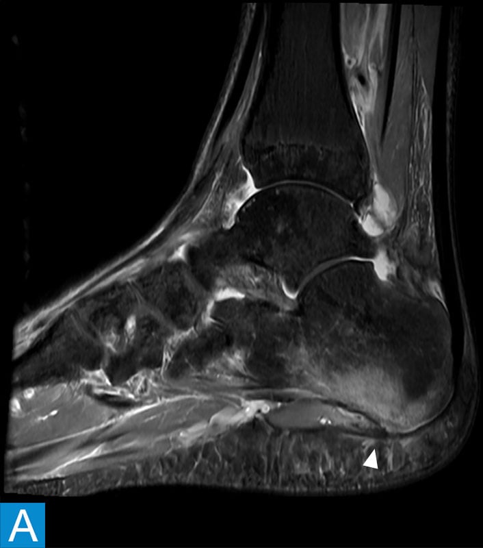
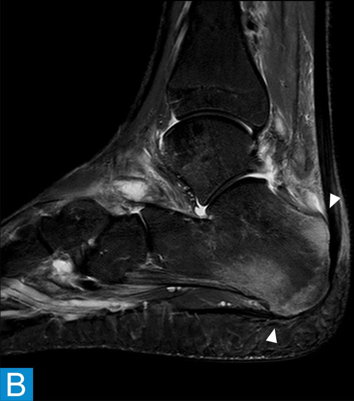
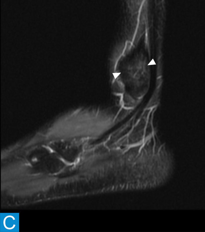
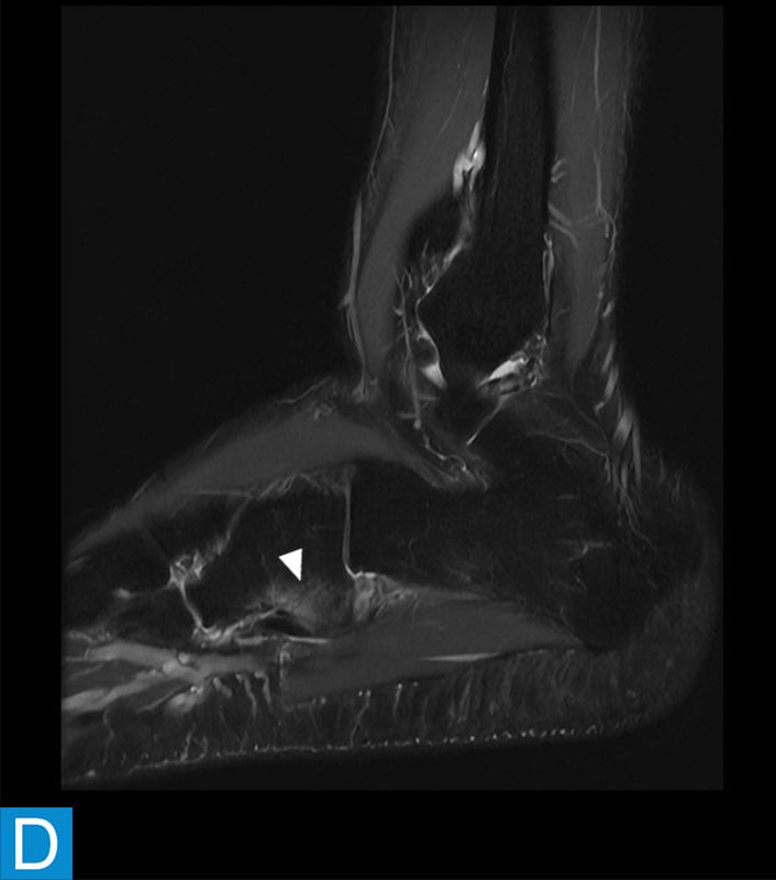
Figure 1. Serial sagittal inversion recovery (STIR) MRI of the right hindfoot demonstrating migratory, entheseal-pattern bone marrow edema (white arrowheads) over four years. (A) At presentation, there is pronounced edema within the calcaneus at the plantar fascia origin. (B) At 6 weeks, MRI shows persistent calcaneal edema with new edema at the Achilles insertion. (C) After initiation of adalimumab, there is new edema within the distal fibula with decreased calcaneal edema. (D) During a flare following medication discontinuation, MRI shows new edema within the cuboid with decreased calcaneal and fibular edema.
Discussion
Bone marrow edema on MRI is a common finding in the evaluation of foot and ankle pain, but its differential diagnosis extends well beyond mechanical stress injury. In young, active patients, calcaneal bone marrow edema is frequently attributed to stress fracture. However, bone marrow edema is nonspecific and can also signify inflammatory enthesitis, infection, or neoplasm (e.g., osteoid osteoma) [1,2]. Enthesitis, inflammation where tendons, ligaments, or fascia insert into bone, can produce an overlapping MRI appearance, including periosteal reaction, marrow edema, and adjacent soft tissue swelling, and is a hallmark of the SpA spectrum, most commonly affecting the calcaneal insertions of the plantar fascia and Achilles tendon [3].
This case presents several clinical lessons. First, the features distinguishing inflammatory enthesitis from stress fracture in this patient included edema centered at tendon and fascial insertions rather than along trabecular stress lines, associated tenosynovitis and periostitis, bilateral and multisite involvement, surrounding soft-tissue swelling, and failure to improve with offloading. Such a combination of findings should prompt re-evaluation of the original diagnosis and consideration of alternative etiologies [4].
Second, the Assessment of SpondyloArthritis international Society (ASAS) classification criteria for peripheral SpA require enthesitis, arthritis, or dactylitis plus at least one SpA feature such as HLA-B27 positivity [3]. This patient met criteria through enthesitis combined with HLA-B27 positivity. ANA positivity is not typically a feature of spondyloarthropathy, and in this case prompted an evaluation for overlap with other systemic autoimmune rheumatic diseases, but these were not diagnosed. Detection of ANA by indirect immunofluorescence at a serum dilution of at least 1:100 has been reported in over 50% of individuals with psoriatic arthritis [5], which is part of this spectrum of “seronegative” SpA. This patient had GI and dermatological symptoms that were potentially relevant given associations of inflammatory bowel disease (IBD) and psoriasis with this spectrum of inflammatory arthritis. Each of these symptoms required specialty evaluation to evaluate further.
Third, tumor necrosis factor (TNF) inhibitors have demonstrated efficacy in peripheral SpA enthesitis. In the ABILITY-2 trial, adalimumab significantly improved enthesitis scores compared with placebo [6], and meta-analyses show anti-TNF, anti-interleukin (IL)-17, and anti-IL-12/23 agents achieve enthesitis resolution with pooled risk ratios of approximately 1.9 versus placebo [7]. This patient’s partial response with flares upon discontinuation is consistent with the chronic, relapsing nature of SpA enthesitis.
When a young patient presents with calcaneal bone marrow edema unresponsive to offloading, clinicians should look beyond stress injury. Serial imaging can be particularly informative: the migration of bone marrow edema to new entheseal sites over time, rather than indicating progressive healing, suggests a pattern more consistent with inflammatory enthesitis than with mechanical overload. Entheseal-pattern edema, tenosynovitis, bilateral involvement, and systemic inflammatory features should prompt HLA-B27 testing and rheumatologic referral to avoid delay of diagnosis.
AI Use Disclosure
We used generative artificial intelligence (OpenEvidence) to assist with literature review, drafting, and formatting of this case report. We reviewed, edited, and approved all content and take responsibility for the final version.
Authors



References
- Schroeder JD, Trigg SD, Capo Dosal GE. Bone stress injuries: diagnosis and management. Am Fam Physician. 2024;110(6):592-600. PMID: 39700362.
- Rios AM, Rosenberg ZS, Bencardino JT, Rodrigo SP, Theran SG. Bone marrow edema patterns in the ankle and hindfoot: distinguishing MRI features. AJR Am J Roentgenol. 2011;197(4):W720-9. doi: 10.2214/AJR.10.5880.
- Rudwaleit M, van der Heijde D, et al. The assessment of SpondyloArthritis International Society Classification criteria for peripheral spondyloarthritis and for spondyloarthritis in general. Ann Rheum Dis. 2011;70(1):25-31. doi:10.1136/ard.2010.133645.
- Expert Panel on Musculoskeletal Imaging; Morrison WB, Deely D, Fox MG, et al. ACR Appropriateness Criteria® stress (fatigue-insufficiency) fracture including sacrum excluding other vertebrae: 2024 update. J Am Coll Radiol. 2024;21(11S):S490-S503. doi:10.1016/j.jacr.2024.08.019.
- Silvy F, Bertin D, Bardin N, et al. Antinuclear antibodies in patients with psoriatic arthritis treated or not with biologics. PLoS One. 2015;10(7):e0134218. doi:10.1371/journal.pone.0134218.
- Mease P, Sieper J, Van den Bosch F, Rahman P, Karunaratne PM, Pangan AL. Randomized controlled trial of adalimumab in patients with nonpsoriatic peripheral spondyloarthritis. Arthritis Rheumatol. 2015;67(4):914-923. doi:10.1002/art.39008.
- Mourad A, Gniadecki R. Treatment of dactylitis and enthesitis in psoriatic arthritis with biologic agents: a systematic review and metaanalysis. J Rheumatol. 2020;47(1):59-65. doi:10.3899/jrheum.180797.