Elbow Arthroscopy
Elbow arthroscopy is a minimally invasive technique used by orthopedic surgeons to diagnose and treat a range of conditions affecting the joint. Arthroscopy of the elbow is less common than that in other joints such as the knee or shoulder. The constraints of working in the elbow, a very tight space in which the nerves pass closely over the joint, require specialized training. Proximity of those nerves also constitutes the major risk specific to the procedure, nerve injury that can result in temporary numbness.
As in other joints, arthroscopy of the elbow involves the use of fiberoptics and a tiny camera that is inserted through small incisions or portals. Magnified pictures from the camera are projected onto a television monitor in the operating suite. When the procedure is employed to treat an injured or diseased joint, the orthopedic surgeon inserts miniaturized surgical instruments through an additional portal.
In contrast to traditional surgery, using large incisions to open the joint, there is no injury to surrounding soft tissues with arthroscopy. Moreover, the technique allows the surgeon to view the elbow joint from multiple angles, allowing for a more thorough evaluation.
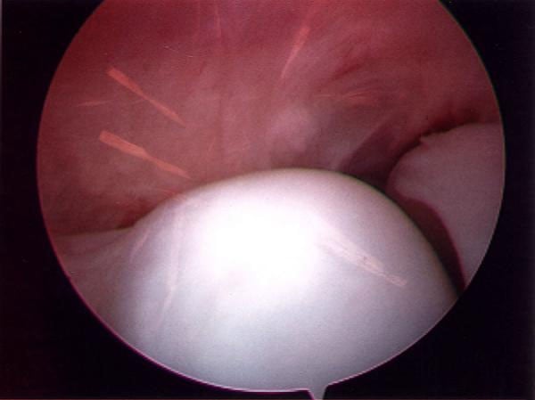
Image of the elbow joint obtained through arthroscopy.
Before performing an elbow arthroscopy, the orthopedic surgeon obtains an MRI (magnetic resonance imaging) and X-rays of the joint. Respectively, these images provide information about the soft tissues surrounding the bones – cartilage, tendons and ligaments – and the bones themselves which may have irregularities including spurs (extra growths that can cause pain and inflammation). Placement of the portals may also be guided by these findings.
Elbow arthroscopy is used as a diagnostic tool for pain, stiffness, and loss of motion in the joint. Some of the more common diagnoses include:
- adhesions, soft tissue bands that block motion as a result of a previous injury to the elbow, such as a fracture
- contractures, a condition in which the muscle and tendons are abnormally contracted, thereby limiting range of motion, loose bodies, fragments of bone or cartilage that break loose causing pain, catching and locking of the joint, and
- arthritis, a disease that is characterized by the wearing away of cartilage, the tissue that helps bones glide smoothly against each other during movement.
An orthopedic surgeon can also treat certain conditions using arthroscopy.
Arthritic bone can be debrided – a technique that renders the surfaces that come in contact with one another smoother and less likely to cause stiffness and catching during movement. Loose bodies can be removed and adhesions and contractures released, thereby improving or restoring range of motion. And, arthroscopy is used by some orthopedic surgeons to debride or cut away the unhealthy portion of the tendon in tennis elbow.
Elbow arthroscopy is an especially useful technique for the diagnosis and treatment of problems in in throwing athletes, especially baseball pitchers. It is used to confirm injuries of the ulnar collateral ligament (UCL) and to treat spurs and cartilage injuries that occur – particularly in the posterior aspect of the elbow.
Elbow arthroscopy is performed under regional or local anesthesia and hospitalization is not required.