Cavus Foot
Medically reviewed by John S. Blanco, MD ; Emily R. Dodwell, MD, MPH, FRCSC ; Shevaun Mackie Doyle, MD ; David M. Scher, MD
The treatment of foot deformities in children is different than in adults. It must preserve the integrity of the growth plates in the bones to allow for their continued growth and development. HSS has pediatric orthopedists who specialize in this field, and cavus foot is one of the pediatric foot deformities they commonly treat.
What is cavus foot?
Cavus foot is a condition in which a person’s foot has an excessively high arch. The condition frequently affects both feet, and it is often progressive. The natural arch in a normal foot helps you maintain balance and absorbs shocks when you walk or run. In high arch feet, more pressure can be exerted on the heel and ball of the foot when walking, causing foot pain. It may make the foot less flexible.
What is cavovarus foot?
A cavovarus foot is a variation of cavus foot deformity, in which not only is the arch elevated (“cavus”) but the foot is also turned inward (“varus”). This deformity can affect the alignment of the foot and may lead to problems with walking, balance, and shoe wear.
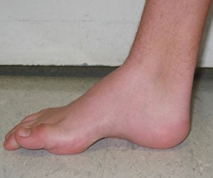
Cavovarus foot – Medial to lateral view of a right cavovarus foot. Note the pathognomonic high arch.
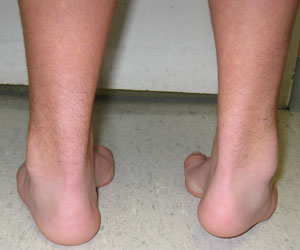
Posterior view of the same patient showing cavovarus foot in both feet.
Cavovarus is commonly associated with clubfoot. However, cavovarus foot that develops over time can indicate the presence of a neurologic problem, the presence of a cyst or, possibly, a tumor in the spinal cord. More commonly, however, it is the result of Charcot-Marie Tooth disease, a hereditary disorder in which the conduction speed of nerves slows over time and causes weakness of the muscles of the hands, lower extremities, and feet. While Charcot-Marie Tooth disease is not life-threatening, the foot problems that accompany it can be disabling. In fact, because other symptoms may be mild or non-existent, the cavovarus foot may be the key to diagnosis.
Another variation of cavus foot, known as calcaneocavus, is characterized by an excessively high arch and an elevated heel (calcaneus bone).
In this condition, the hindfoot (the back part of the foot, including the heel) is in an overly extended or dorsiflexed position, while the forefoot is plantarflexed (pointed downward). This creates a significant angle between the front and back parts of the foot, contributing to the high arch.
What causes cavus foot?
Cavus foot can be congenital and idiopathic (appearing at birth with no known cause) or acquired as a result of other conditions, including neurological disorders (such as Charcot-Marie-Tooth disease, spina bifida, tethered cord syndrome, cerebral palsy or a stroke), spinal tumors, peripheral nerve injuries, muscle imbalances caused by muscular dystrophy, polio or other conditions, inflammatory diseases such as rheumatoid arthritis, or genetic conditions such as Freidreich's ataxia.
What are the symptoms of cavus foot?
Because the foot is not properly aligned, the uneven weight distribution can cause pain when standing or walking for long periods of time. Metatarsalgia (pain in the forefoot), calluses, and/or corns may develop. Patients are also prone to ankle sprains or even stress fractures of the ankle or metatarsals.
Other conditions are also caused by or commonly occur in conjunction with cavus foot. These include plantar fasciitis, hammertoes, claw toes, ankle instability, and stress fractures of the metatarsals.
What is the treatment for cavus foot?
In mild cases, an orthopedist may recommend use of orthopedic shoes or orthotics (shoe inserts that can help evenly distribute weight across the foot) and routine surveillance for progression of the problem. Physical therapy may also help. More severe cases or when cavus foot is associated with other medical problems, surgery is often needed.
What is the surgery for cavus foot?
Surgery for cavus foot may be a combination of osteotomies, in which the bones are cut and repositioned, and soft tissue procedures. Ligaments may be released and tendons transferred to help rebalance and supplement weakened muscles in the foot and leg. There is no single procedure or even group of procedures. Each case is different, but the goal is the same: to realign the foot and ankle joints, correct the deformity, and rebalance the soft tissues and muscle forces.
Cavus foot soft tissue surgeries
Stretching forces from various soft tissues such as muscles, tendons, and/or the plantar fascia may be partly responsible for the high arch. The surgical release of one or more of these structures, or a tendon transfer may help correct the deformity. These may sometimes be performed alone but more often are done in conjunction with bone osteotomies and/or joint fusions.
Osteotomies
Depending on each patient’s case, osteotomies (cutting and realignment of bones) may be performed on the first metatarsal, bones of the midfoot, and/or the calcaneus (heel bone).
Joint fusions
Fusion surgeries can reduce pain and improve the stability of the foot by eliminating motion in the affected joints. In a triple arthrodesis surgery, the subtalar joint, talonavicular, and calcaneocuboid joints of the hindfoot are fused.
What is the recovery time for cavus foot surgery?
Most cavus foot surgery recovery takes three months, accounting for post-operative healing and rehabilitation. Depending on the procedures needed to realign the foot, some recoveries are quicker than others. Generally, osteotomies and tendon transfers heal in six weeks while the limb is immobilized. Physical therapy is then needed for another 6 to 8 weeks to reduce the stiffness that ensues after extremity immobilization and have the redirected muscle forces work in concert with the improved alignment.
What are the risks or complications of cavus foot surgery?
The lower leg needs to be immobilized and free from any weightbearing activity for 6 to 8 weeks after surgery, therefore patients are at risk for deep venous thrombosis. We take precautions to avoid this by using aspirin or other blood-thinning medications after surgery. Other risks include nonunion of the osteotomies and rupture of tendon transfers. However, these are rare occurrences. Additionally, as with any surgical procedure, there is the risk of infection, but patients are thoroughly treated with external and intravenous antimicrobial agents to mitigate this worrisome risk.
Explore Related Patient Stories
View All Patient Stories
Ryan Bailie Plaza
Maplewood, NJ
Cavus Foot