Micro-Computed Tomography Core
Introduction
The micro-computed tomography (microCT) core provides high-resolution assessments of density, geometry and microarchitecture of mineralized tissues, such as bones and teeth, calcification as result of pathology, or soft tissues and biomaterials stained with radiographic contrast media. Studies performed by the HSS microCT core include:
- density and architecture analysis of human biopsies (Fig.1)
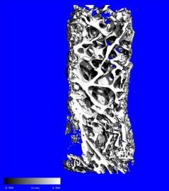
Figure 1: Human iliac crest biopsies
- phenotype characterization of bones and teeth in animal models (Fig. 2)
Adult mouse femur Mouse knee joint 7 month-mouse molar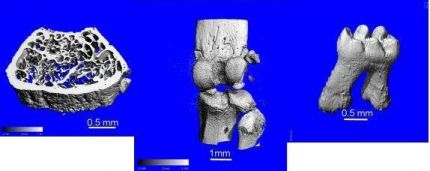
Figure 2: Phenotype characterization of bone and teeth
- studies of particle-induced osteolysis (Fig. 3)
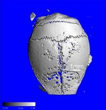
Figure 3: Mouse calvaria: particle-induced osteolysis
- characterization of dystrophic calcifications
- visualization of bone implants
- biomaterial characterization
- vascular mapping
Equipment
Scanner: Scanco Medical MicroCT 35 (Fig. 4)
- Resolution: 1.75-38 microns. In a typical mouse bone characterization the resolution used is 6µm (for femur/tibia) or 10µm (for knee).
- Wide range of KVp (45-70)/selection of different filters, allowing for high- (soft/developing mineralized tissue), normal- (mature bone) and low-contrast (implants, tooth enamel) scans.
- Incremental scanning: Every field of view chosen for analysis is scanned by increments using a 2048 x 2048 CCD detector. This enables a precise choice of the analyzed area by the user.
- Batch scanning: Multiple specimens are loaded for scanning with pre-selected settings for efficient use of the system.
- Different setups for analysis of variable size samples
- Reproducibility of analysis: % range of measurements from repeated scans at a 6µm resolution:
- calibration phantom: <0.2% for mineral density
- mouse femur trabecular bone: <0.5% for mineral density, <0.7% for trabecular bone volume fraction, relative bone surface, trabecular thickness, number, and separation
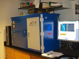
Figure 4: Scanco Medical MicroCT 35
3D reconstruction and processing: HP Integrity server workstation.
The Scanco workstation combined with the 64bit-software for densitometry and histomorphometry analysis provides:
- archiving of scans
- fast 2D and 3D evaluation of reconstructed volumes
- automated density scaling correction
- automated beam hardening and center of rotation correction
- versatile definition of volume contour for analysis by user
- handling of very large datasets
- complete array of direct morphometry measurements in 3D
Core Support
The microCT core-associated staff meets with all new users before the beginning of analyses to discuss the technical options for the users’ purposes. The focus is on an efficient use of the system within the study budget. A limited amount of pilot analyses is also available for first-time studies.
Fee Schedule
- Scanning:
$75/hour (NIH-funded projects)
$100/hour (academic, non NIH funded)
$200/hr (corporate) - Analysis:
$75/hour (NIH-funded projects)
$100/hour (academic, non NIH funded)
$200/hr (corporate or other) when provided by the Core or $25/hour on the Core workstation by the user (training on analysis software is provided to users at the hourly rate).
Typical scanning time for mouse femurs: 6-10µm resolution ~1.5h (4 femurs can be scanned in parallel).
To schedule samples please review the Service Request Process and complete the form as provided below:
Service Request Process
Service Request Form
Contact Information
Kyung Hyun Park-Min, PhD
Assistant Scientist and Director
Email: ParkminK@HSS.edu
Hayat Benlarbi
Senior Research Technician
Email: BenlarbiH@HSS.
Phone: 212-774-7329