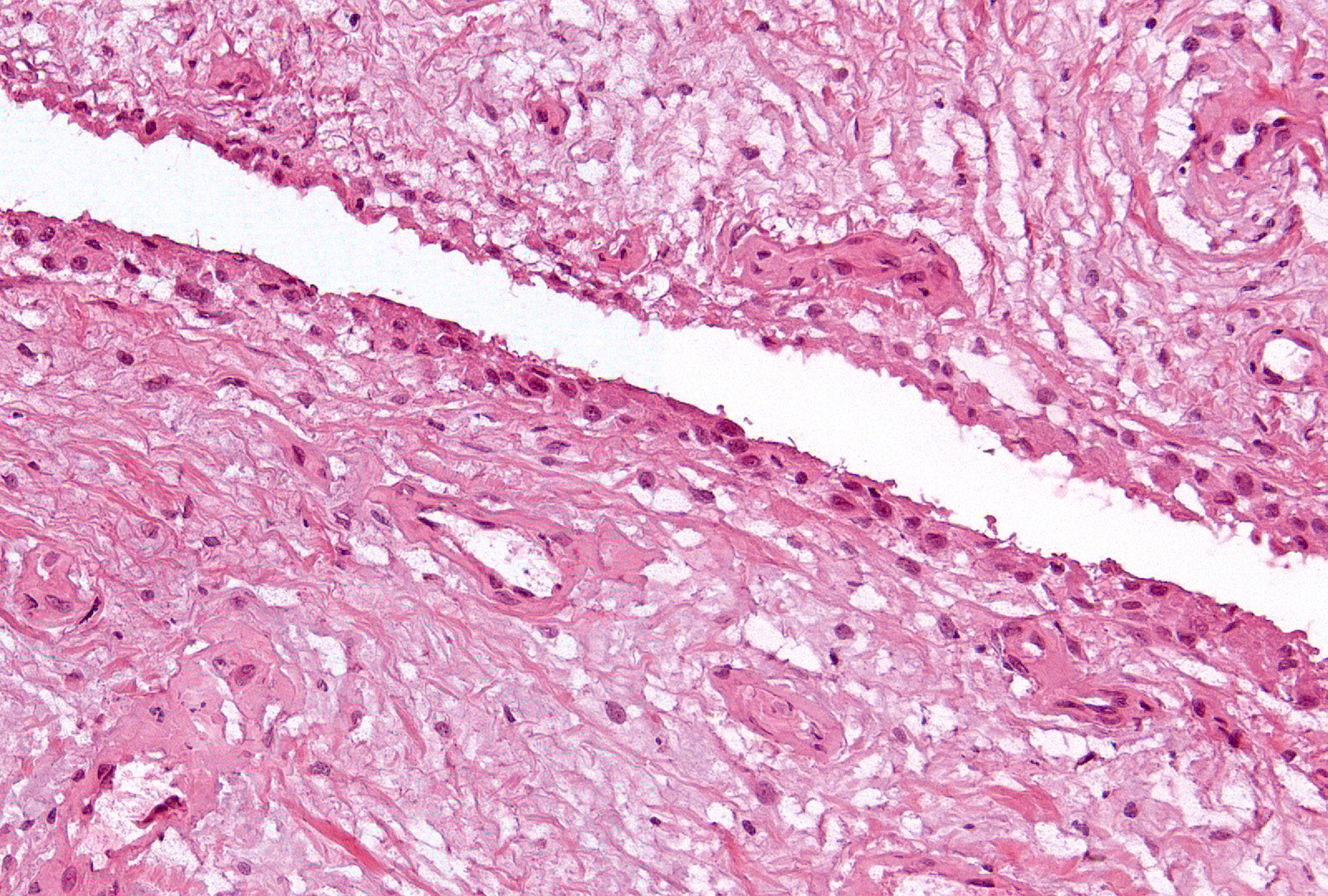
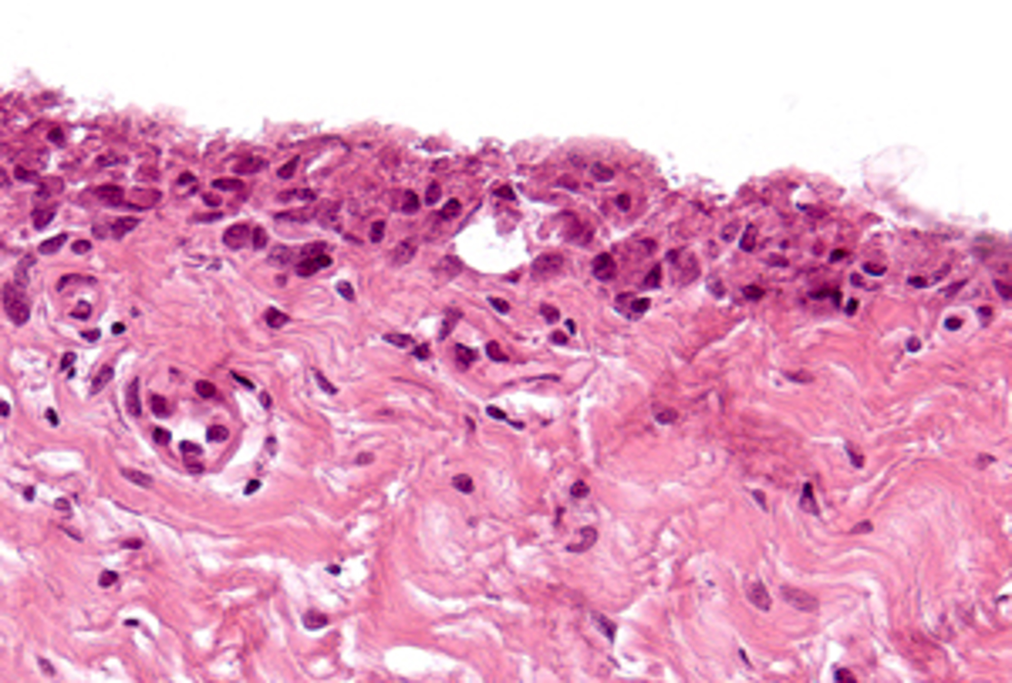
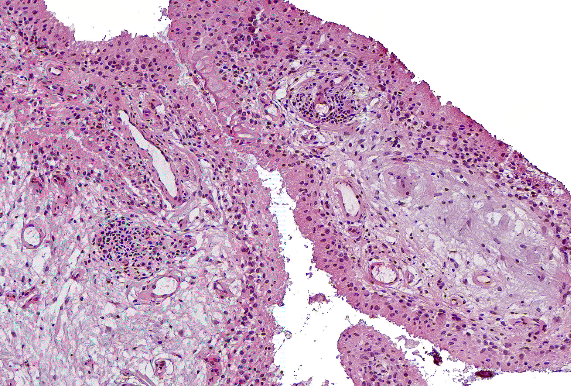
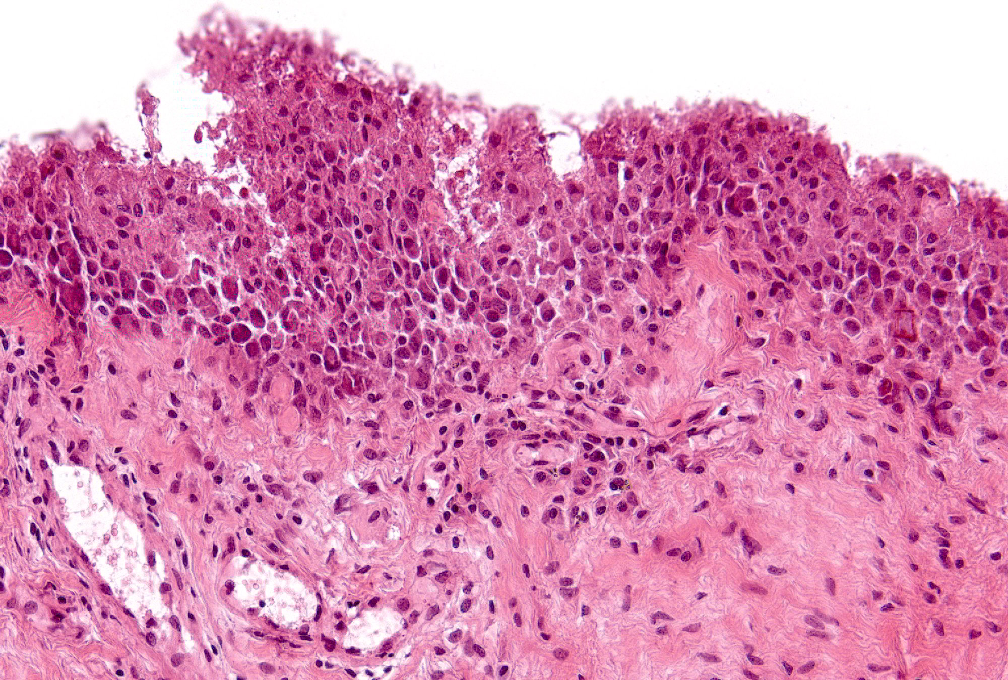
Synovitis Pathology
Synovial Lining Hyperplasia
Feature:
Number of cells that comprise the height of the synovial lining layer seen in over half the identifiable synovial surfaces (Exclude sectioning artifact that results in “thickening” of the lining due to tangential cutting.)
Magnification: 4-5x
Grading:
- 0: (6%) 1 cell thick
- 1: (43%) 2-3 cells thick
- 2: (31%) 3-4 cells thick
- 3: (20%) > 4 cells thick
Grade 1 | Grade 2 | Grade 3
Back to Pathology of Synovitis