Fracture Dislocations & Treatment
Case Example
Acetabular Fracture with Posterior Hip Dislocation
A 35-year-old female fell from a standing height and felt an immediate onset of severe right-sided hip pain. She was taken to a local hospital where x-ray and CT scan evaluation revealed a right-sided Posterior Wall acetabular fracture and associated posterior hip dislocation. The local medical team then contacted David L. Helfet, MD at the Orthopedic Trauma Service of Hospital for Special Surgery and a patient transfer was arranged following placement of skeletal traction. Open Reduction and Internal Fixation (ORIF) was then performed with placement of 3 spring plates along the posterior wall fragment and an anatomic reduction was obtained. She returned for regular follow-up and healed uneventfully and at her latest follow-up visit at 5 months she presents with excellent radiographic and clinical results including a healed acetabular fracture, complete resolution of hip pain, full range of hip motion and return to her pre-injury activities.
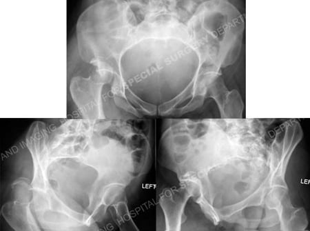
Anteroposterior and Judet radiographic views (Obturator Oblique and Iliac Oblique views) revealing
a right-sided Posterior Wall acetabular fracture and associated posterior hip dislocation.
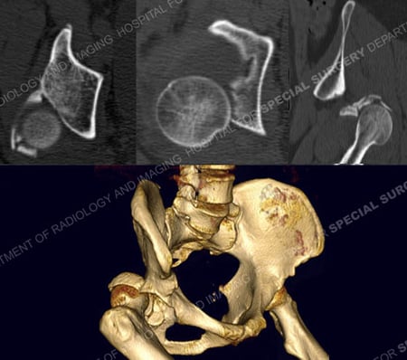
CT scan images including 3D CT reconstruction image further delineating the fracture pattern.
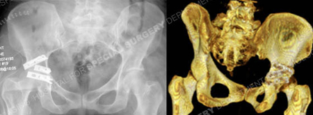
Postoperative anteroposterior and 3D CT reconstruction image revealing an acceptable reduction and placement of hardware.
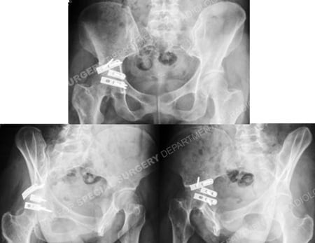
Anteroposterior and Judet radiographic views (Obturator Oblique and Iliac Oblique views)
at 5 months illustrating a healed Posterior Wall acetabular fracture.
Research Publications
The HSS Orthopedic Trauma Service has conducted many studies. Please see our publications on acetabular fractures.