Radius Fractures
Case Example
A 14-year-old male was hit in the right forearm while playing football and experienced immediate pain and swelling. He was taken to a local hospital and radiographs revealed a closed right-sided distal radius fracture and associated dislocation of the distal radius ulna joint (Galeazzi fracture-dislocation). He was referred to David L. Helfet, MD at the Orthopedic Trauma Service of Hospital for Special Surgery for definitive management. Open reduction and internal fixation (ORIF) was performed and the fracture was reduced and fixed using a locking plate and screws including an interfragmentary lag screw. He returned for regular follow-up and healed uneventfully. At the time of his latest follow-up visit, 8 months following fracture surgery, he has excellent radiographic and clinical results including a healed distal radius fracture in excellent alignment, resolution of pain, full range of motion, and a return to pre-injury activities.
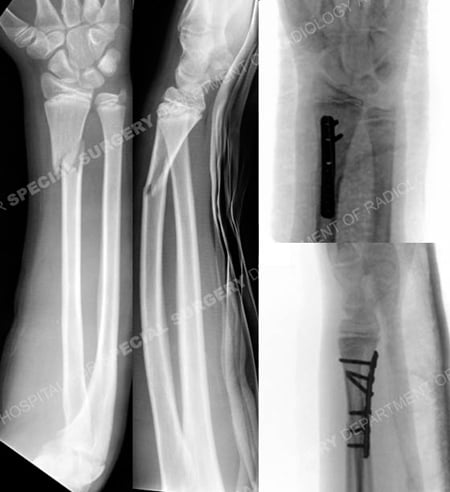
Anteroposterior and lateral radiographs (left images) revealing a right-sided distal radius fracture and associated dislocation of the distal radius ulna joint (Galeazzi fracture-dislocation) and intraoperative fluoroscopic radiographs (right images).
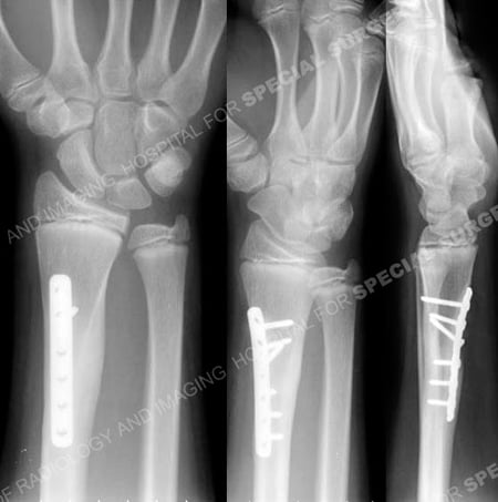
Anteroposterior and lateral radiographs 8 months following fracture surgery illustrating a healed distal radius fracture in excellent alignment.
Research Publications
The HSS Orthopedic Trauma Service has conducted many studies. Please see our publications on radius fractures, fractures in adolescents, and wrist fractures.