Foot Fractures
Case Example
A 68-year-old male was involved in a motor vehicle accident when the car he was driving was struck from behind and pushed into the vehicle in front of him. He was brought to the HSS Orthopedic Trauma Service with complaints of left foot pain and radiographs revealed a left-sided, displaced, intra-articular calcaneus (heel bone) fracture with a depressed articular segment. Fracture surgery was performed by Dr. David L. Helfet using a minimally invasive technique with elevation of the depressed segment, reduction and fixation of the posterior facet and tuberosity including interfragmentary lag screws. He returned at regular follow-up intervals and healed uneventfully, and at 6 months following surgery he presented with good radiographic and clinical results including a healed calcaneus fracture, significant improvement of pain symptoms, and a return to his prior activities of daily living.
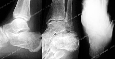
An injury radiographs revealing a calcaneus fracture.
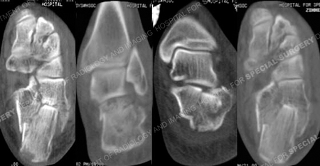
CT scan images further delineating the fracture pattern.
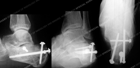
Radiographs 6 months following fracture surgery revealing a healed calcaneus fracture in excellent alignment.
Research Publications
The HSS Orthopedic Trauma Service has conducted many studies. Please see our publications on foot fractures and minimally invasive fracture treatment.