Pachydermodactyly, a Rare and Benign Digital Fibromatosis, Diagnosed in a 15-Year-Old Boy
From Grand Rounds from HSS: Management of Complex Cases | Volume 8, Issue 3
Case Report
A 15-year-old boy presented with a 5-year history of bilateral swelling of the proximal interphalangeal (PIP) joints. The swelling had occurred gradually, until he had visible deformity of both hands. He denied pain or stiffness in his fingers and could write and play sports without limitation. He had no other symptoms, including pain or swelling in his other joints, morning stiffness, fever, or rash. He denied any activity causing repetitive trauma to his fingers, although he did admit to the habit of cracking his knuckles multiple times a day. He had no significant medical or family history. He had previously seen 3 hand surgeons and a pediatric rheumatologist, with no diagnosis.
Clinical examination revealed bilateral periarticular swelling of the PIP joints of the second through fifth digits, with no joint tenderness or decreased range of motion. He had areas of thickened skin on the fingers of both hands (Fig. 1).
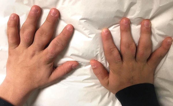
Figure 1: Bilateral swelling of the PIP joints, with thickening of the overlying skin.
There were no other joint findings, and his physical examination was otherwise normal. Laboratory investigations, including complete blood count, C-reactive protein, erythrocyte sedimentation rate, antinuclear antibody testing, rheumatoid factor, anti-cyclic citrullinated peptide antibodies, and human leukocyte antigen B27 were all normal. X-ray showed soft-tissue swelling around the PIP joints, with normal joint spaces, articular surfaces, and bone morphology (Fig. 2).
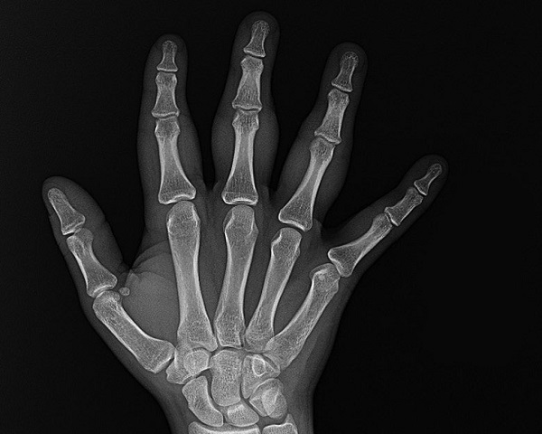
Figure 2: X-ray showing soft-tissue swelling around the PIP joints, without joint space narrowing, erosions, or other bony abnormalities.
Magnetic resonance imaging showed soft-tissue prominence, including collateral ligament thickening with infiltration of subcutaneous fat, but no effusions or erosions. In the absence of clinical, laboratory, and radiological findings suggestive of an inflammatory arthropathy, a diagnosis of pachydermodactyly (PDD) was made. The patient and his family were educated on the nature of this disease, and he was encouraged to refrain from repetitive mechanical trauma to his fingers, including knuckle cracking.
Discussion
PDD is a rare, benign, acquired digital fibromatosis marked by painless, progressive swelling of the periarticular soft tissues of the fingers, without joint involvement. Adolescent males are most commonly affected [1]. Although the precise etiology remains unknown, the disorder is thought to be related to repetitive minor mechanical trauma, including frequent cracking of the finger joints [2]. Patients present with progressive, painless swelling around the PIP joints, often with thickening of the overlying skin. PDD is diagnosed in patients with typical examination features and the absence of findings consistent with inflammatory arthropathy on imaging and laboratory tests. [1]. Although biopsy is not routinely necessary for diagnosis, histopathology shows hyperkeratosis, acanthosis, and dermal thickening, with increased collagen bundles and fibroblasts [3]. The prognosis is benign, and treatment is not required unless patients are disturbed by the appearance of the fingers. Intralesional corticosteroid injection and surgical excision have been used with benefit in some cases; avoidance of mechanical trauma is recommended [4].
PDD can mimic the appearance of an inflammatory arthropathy, such as psoriatic arthritis, rheumatoid arthritis, or juvenile idiopathic arthritis. Patients with PDD are often misdiagnosed, leading to unnecessary concern and treatments. Careful attention to the physical exam, history, and imaging is crucial in order to make the correct diagnosis. Awareness of this clinical entity allows physicians to provide reassurance to their patients and to avoid prescribing unwarranted antiinflammatory or immunosuppressive treatment.
Posted: 10/1/2019
Authors
Halide Ozge Basaran, MD
Visiting Research Fellow

Attending Orthopedic Surgeon, Hospital for Special Surgery
Assistant Professor of Orthopedic Surgery, Weill Cornell Medical College

Attending Physician, Hospital for Special Surgery
Assistant Professor of Pediatrics, Weill Cornell Medical College
References
- Dallos T, Oppl B, Kovács L, Zwerina J. Pachydermodactyly: a review. Curr Rheumatol Rep. 2014;16(9):442.
- Rancy SK, Granstein RD, Bansal M, Barley CL, Fields TR, Wolfe SW. Pachydermodactyly: a case report including histopathology. J Hand Surg Am. 2016;41(8):e243–246.
- Naviglio S, Mazzolai M, Ventura A. Bilateral finger swelling in an adolescent. J Pediatr. 2017;1(188):299.
- Jhu H, Song J, Kim do Y. Pachydermodactyly: a benign cutaneous condition that may be misdiagnosed as a joint disorder. J Rheumatol. 2016;43(8):1615–1616.