- HSS Florida

Radiology and Imaging
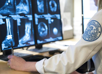
The radiologists at HSS Florida provide expert imaging services to people with soft-tissue injuries and conditions. These doctors have special training in analyzing and interpreting musculoskeletal imaging.
Among the services offered at HSS Florida are MRI, ultrasound and X-ray. Our radiologists have access to the most advanced imaging technologies, such as dynamic ultrasound techniques as well as high-resolution MRIs. Our unparalleled expertise and access to highly advanced technology can help you get the right diagnosis and ensure the most-effective treatment plan possible.
Magnetic Resonance Imaging (MRI)
Magnetic resonance imaging is a diagnostic imaging technology used to detect problems in both bone and soft tissues. MRI depicts soft-tissue injury and abnormalities with greater sensitivity and specificity than conventional imaging techniques.
The MRI technology at HSS Florida incorporates high-spatial resolution and thin-slice imaging. These state-of-the-art techniques (not always available from other healthcare providers) provide highly detailed information about cartilage, tendons and other soft tissues. This helps HSS radiologists make the most precise diagnosis possible.
In addition, we use special techniques to detect cartilage damage in its early stages as well as to obtain more accurate readings for those who’ve already had a joint replacement. Our high-resolution MRI exams do not require invasive intra-articular gadolinium contrast.
Ultrasound
Ultrasound uses high frequency sound waves to produce images. Sound waves are sent and received through a small handheld device known as a transducer. The returning sound waves are used to produce the images. New ultrasound technology allows us to look in great detail at the specific soft tissue structure being examined and at the blood flow in vessels within the soft tissues.
At HSS Florida, ultrasound is also used to guide injections for the delivery of medication (corticosteroids) and other agents into joints, tendons, bursas, cysts, ganglions and neuromas. Because ultrasound images are produced in real time, ultrasound can enhance these procedures by allowing the radiologist to continuously monitor the positioning of the injection.
X-ray Scans (Radiographs)
X-rays, or radiographs, are medical images that are taken using beams of radiation to create a picture on film, based on the different densities of the body’s structures. Radiographs are a reliable and accurate means of obtaining information to help your doctor determine the cause of your pain.
An X-ray examination is commonly used to diagnose a bone fracture, joint malalignment, arthritis, or cause of other painful conditions. HSS uses an array of customized approaches to obtain an optimal view of relevant bones and joints structures.