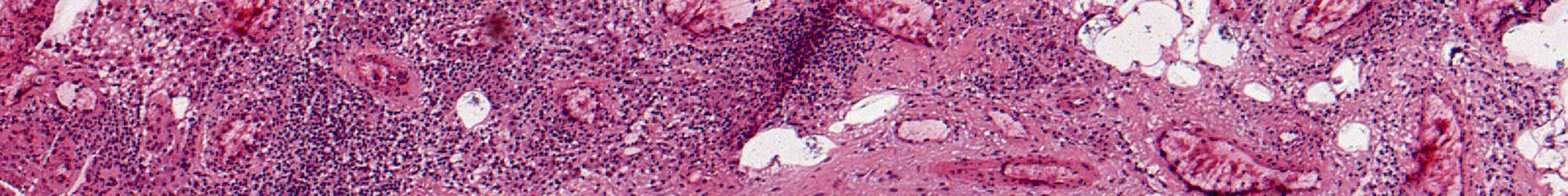
Synovitis Pathology
Synovitis slider
Grading Method for Synovitis Pathology
Welcome to the HSS synovial pathology scoring image directory. Here you will find instructions for scoring 20 histologic features of synovial frozen sections that predict whether the sample synovium is low-, mixed-, or high-inflammatory subtype.
In order to score these samples it is important to carefully select the tissue as presented in the publication. Simply put, the surface of the tissue should be dull and granular, brown or beefy, and the delicate vascular pattern typical of normal synovium should be obscured.
The images are organized into several related categories that were assessed in this study. Clicking on the feature name in this directory will open additional links to representative examples that can be further inspected and magnified.
You can enter the 10 individual histology feature scores of a given sample to generate a single, summary, histology prediction score.
Approach to Grading
Grading requires staining with Harris modified Hematoxylin and Eosin. These features are assessed using frozen sections.
The slide should be previewed at low magnification to assess the adequacy of the tissue as well as overall orientation of the tissue on it.
The various features are to be graded at specific magnifications, with or without verification using higher magnifications.
The criteria and magnification for grading each feature are defined in this directory.
In general, the criteria for the various grades are met when they are present in 2 noncontiguous fields at the appropriate magnification.
If the criteria for each higher grade are not met, then the assigned grade is the lowest that can still be met.
Inflammation
Surface Features
Stromal Features
Specific Cytology
Features not illustrated: Granulation Tissue, Necrosis, Infarction, Synovial Chondrometaplasia and Eosinophils