Revolutionizing Spinal Imaging — without Radiation
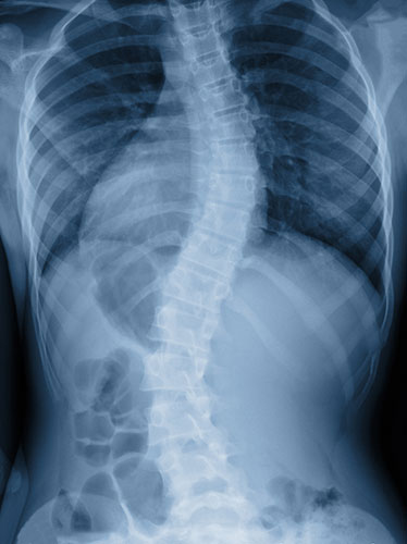
For years, experts have sought new and innovative methods to minimize radiation exposure for adolescent idiopathic scoliosis patients, who typically require between 10 and 25 X-rays over the course of their treatment to assess skeletal alignment and curve progression. In 2013, HSS took a major step toward that goal with the introduction of EOS imaging, an ultra-low-dose X-ray system that scans patients in a standing position. While the technology has dramatically reduced radiation exposure in these patients versus traditional X-rays, in 2019 HSS embarked on implementing a novel method that does not require ionizing X-ray radiation.
The initiative will focus on refining the use of 3D surface imaging technology to map skeletal alignment in scoliosis patients and has brought together more than 20 different experts from across HSS, including those from pediatric orthopaedics, motion analysis and radiology, as well as scientists from Technion Israel Institute of Technology.
The imaging process used by the technology, called 3dMD, is known as stereophotogrammetry. A patient stands in a small room while 30 high-resolution cameras capture images of their body. "The images are extremely precise, with sub-millimeter accuracy," says Howard Hillstrom, PhD, Director of the Leon Root, MD Motion Analysis Laboratory at HSS. "We may also capture this 3D topographic data for slow movements, such as forward bending, at 10 frames per second." The overarching goal is to feed the observed surface topography into a machine learning algorithm to estimate spinal alignment in a variety of poses — without any radiation.
"Unlike EOS imaging, which takes 5 to 10 seconds to capture an image, with 3dMD it takes only two thousandths of a second to get a 3D assessment of the patient’s entire body, from the top of their head to the bottom of their toes," says Roger Widmann, MD, Chief of the Pediatric Orthopaedic Surgery Service at HSS. While the technology has been used for years for cranial facial reconstruction, HSS is leading the development and implementation of highresolution scanning in orthopaedics.
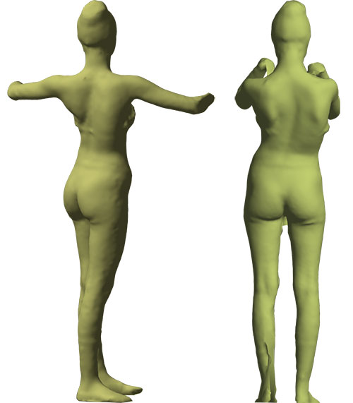
These surface reconstruction images were created by the 3dMD system, showing a patient in a torso twist, with arms elevated at a 45-degree angle, and in the standard posture for EOS imaging.
Before 3dMD can be adopted as the primary screening tool in scoliosis patients, the team of researchers at HSS and Technion must develop and validate surface topography– based parameters that can reliably estimate spinal alignment and body symmetry, says Dr. Hillstrom. "There’s sometimes a significant discordance between internal anatomy — which can be seen on an X-ray or CT scan — and how the spine looks from the outside," explains Dr. Widmann.
HSS has started a registry to track patients who have undergone both 3dMD scans and EOS studies — data that can be used in conjunction with clinical and patient-reported outcomes measures so that researchers can begin to develop and validate clinically relevant and patient-centered parameters. The end goal is to enroll 2,000 patients. "It’s going to take a lot of data to draw accurate conclusions," Dr. Widmann says. "We do have a specific question here, whether we can accurately and reliably correlate 3D skeletal anatomy with surface topography. There are also many other interesting questions that you can answer with such a huge data set."
Down the line, Dr. Widmann hopes to not only expand use of the 3dMD system among adult spine and arthroplasty surgeons at HSS, but also to explore using it in other innovative ways, such as in combination with motion capture technology to analyze patients’ walking patterns and joint movements. Another possibility may be to use it for scanning residual limbs for prosthetics, instead of using plaster casting. "The 3dMD system has the potential to become a standard imaging modality in orthopaedic surgery," says Dr. Widmann. "Here at HSS, we have to prove it, so that’s exactly what we’re doing."
Video - Revolutionizing Scoliosis Imaging Without Ionizing Radiation
Back to HSS Annual Report 2018-19