Osteolysis
What is osteolysis?
Osteolysis is a progressive condition where bone tissue is destroyed. In this process, bones lose minerals (mostly calcium), softens, degenerates and become weaker.
What causes osteolysis?
Osteolysis occurs when cells in the bone called osteoclasts increase their activity and break down the surrounding minerals. There are different types of osteolysis, and each has specific mechanisms that lead to this increase in osteoclast activity and the resulting condition of demineralization.
What are the different types of osteolysis?
The most common types of osteolysis, which are generally unrelated to one other, include:
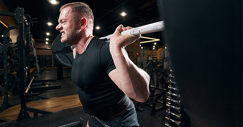
- distal clavicular osteolysis – commonly called “weight lifter’s shoulder”
- periprosthetic osteolysis – which affects some people who have had joint replacement surgeries
- acro-osteolysis (rare) – in which bone in the distal phalanges (fingers or toes) of the hand or feet erode and degenerate
Distal clavicular osteolysis: What is weight lifter's shoulder?
Distal clavicular osteolysis – also known as osteolysis of the shoulder (distal clavicle), AC joint osteolysis or “weight lifter’s shoulder” – affects the acromioclavicular joint (AC joint), at the top of the shoulder. This progressive condition destroys bone tissue in the clavicle. The AC joint is where the acromion – a part of the scapula (shoulder blade) meets the clavicle (collarbone) at its distal (outer) end. They are connected by the acromioclavicular ligament. The connection by this ligament enables you to raise your arm above your head. In some people, the distal end of the clavicle at the AC joint can begin to lose calcium, soften and erode.
Osteolysis of the AC joint is most common among weight lifters and other athletes who do significant weight training. But it can also affect people who frequently lift heavy objects overhead (such as construction or factory workers) or otherwise exert force on the shoulder by repetitive overhead movement (such as tennis or squash players, competitive swimmers, etc.).
What causes weight lifter's shoulder?
The specific cause of distal clavicular osteolysis is not fully understood. However, it is thought to be caused by one or a combination of predisposing factors, such as:
- repetitive injuries to the AC joint or distal clavicle
- repetitive motions with heavy weights (hence the name “weight lifter’s shoulder”)
- a combination of the long-term wear on the shoulder joint combined with some predisposing joint condition, such as rheumatoid arthritis
- other underlying diseases or chronic conditions that can affect the AC joint, such as infections
- (rarely) a single blunt force trauma to the clavicle (impact such as by a fall or physical blow)
Among weight lifters, it is thought that certain activities overload the joint, causing microtrauma where the damage doesn’t have time to heal between lifting sessions. This leads the bone to dissolve rather than heal.
What are the symptoms of weight lifter's shoulder?
Key symptoms of this condition include:
- sharp pain in the AC joint or collarbone during activity
- continued dull aching or tenderness of these same areas during inactivity
- inflammation (swelling) in the shoulder or collarbone area
Distal clavicular osteolysis tends to progress rather slowly and starts with dull pain, tenderness, or stiffness in the shoulder that worsens over time. The pain typically presents in the anterior region of the shoulder at the AC joint, and becomes worse with activities that involve heavy lifting, pushing, or throwing. Over time, up to approximately three centimeters of bone may erode.
How do I know if I have weight lifter's shoulder?
The diagnosis of distal clavicle osteolysis can be usually made by physical examination, although imaging tests may be used to confirm the diagnosis or rule out other causes of shoulder pain.
What is the treatment for weight lifter's shoulder?
Treatment is usually nonsurgical, and includes icing and resting the shoulder, taking anti-inflammatory medications, and undergoing physical therapy. In some instances, surgery may be needed. Treatment focuses on reducing pain and minimizing activities that exacerbate the condition while allowing time for the bone to rebuild (remineralization).
Standard conservative treatment includes:
- rest
- icing
- NSAIDs (nonsteroidal anti-inflammatory drugs)
It is also recommended that patients who are smokers quit to help with the bone remineralization process (this means the restoring of calcium in the bone). It may take several months for the bone to recover.
What is the surgery for weight lifter's shoulder?
If conservative treatments do not effectively remineralize the clavicle, surgery to remove part of the end of the clavicle may be required to alleviate symptoms.
Periprosthetic osteolysis
A second type of osteolysis can occur as a complications of joint replacement surgery. Most patients recover from joint replacement surgery with no complications. But occasionally, polyethylene or other materials in a joint implant can wear down. When this happens, debris can accumulate in the surrounding joint tissue. This, in turn, causes inflammation that can result in degeneration of the bone.
In people who have had hip, knee or other joint replacements, a key sign of periprosthetic osteolysis is an aseptic loosening of the joint prosthesis (that is, a loosening of the implant without any indication of infection).
This condition often causes no symptoms until very late, after there has been extensive bone loss. For this reason, joint replacement patients should have periodic follow-up X-rays of their joint. When they do occur, symptoms of osteolysis around a joint prosthesis are generally related to the associated loosening of the implant. They include:
- pain
- weakness
- Stiffness
If you have had a joint replacement, and after time begin experiencing the above symptoms, your surgeon may first order tests, X-rays and MRI imaging to see if you have a postsurgical infection which, separate from periprosthetic osteolysis, can also lead to joint pain and loosening of an implant.
If caught early, various treatments may be used to treat the inflammation and prevent further osteolysis. If bone loss is deemed extensive, the treatment may require surgery to revise the joint replacement (for example hip revision or knee revision).
Acro-osteolysis
Acro-osteolysis is where bone in the distal phalanges (fingers or toes) of the hand or feet erode and degenerate. Osteolysis may be caused by an underlying inflammatory condition. This can include infections, genetic disorders and problems with the endocrine system. Acro-osteolysis is seen frequently in patients who have certain underlying rheumatic and inflammatory conditions, including:
Acro-osteolysis is also found in people who have:
- experienced extensive use of vibratory power tools (such as pneumatic drills)
- been exposed to vinyl chloride
- elevated levels of parathyroid hormone
Acro-osteolysis, in some cases (such as in people who have inflammatory conditions such as scleroderma or severe Raynaud’s disease), may be accompanied by an associated condition known as digital ischemia. This is where the tissue is not getting enough blood flow, and there can be death of skin cells which could lead to ulcerations of the extremities. In other cases, acro-osteolysis may not be associated with digital ischemia.
The most prevalent symptom is pain in the fingers or toes. If there is associated digital ischemia, there can also be color change or breaks in the skin. X-rays are the standard method used to diagnose acro-osteolysis.
Treatment for acro-osteolysis mainly relates to treating the underlying condition. In cases where there is both acro-osteolysis and decreased blood flow to the extremities, local wound care and treatments that can increase blood flow to the extremities can be used.
Osteoloysis research articles for patients
Osteolysis Patient Stories
References
- Miller TT. Imaging of hip arthroplasty. Eur J Radiol. 2012 Dec;81(12):3802-12. doi: 10.1016/j.ejrad.2011.03.103. Epub 2011 May 6. PMID: 21530121.
- Inoue K, Nakano S, Zhao B. Osteoclastic microRNAs and their translational potential in skeletal diseases. Semin Immunopathol. 2019 Sep;41(5):573-582. doi: 10.1007/s00281-019-00761-4. Epub 2019 Oct 7. PMID: 31591677; PMCID: PMC7027942.
- Purdue PE, Koulouvaris P, Potter HG, Nestor BJ, Sculco TP. The cellular and molecular biology of periprosthetic osteolysis. Clin Orthop Relat Res. 2007 Jan;454:251-61. doi: 10.1097/01.blo.0000238813.95035.1b. PMID: 16980902.
Updated: 10/28/2019