Advice to improve your movement, fitness, and overall health
from the world's #1 in orthopedics.
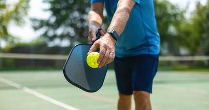
Pickleball injuries are on the rise. Learn more about how to prepare for the game to stay injury free.

With a few straightforward adjustments, you can lessen the impact of golf on your hips and knees.

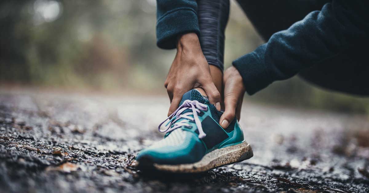
If running a long-distance race is a future goal, the time to start planning is well in advance.
Register for a free virtual workshop!
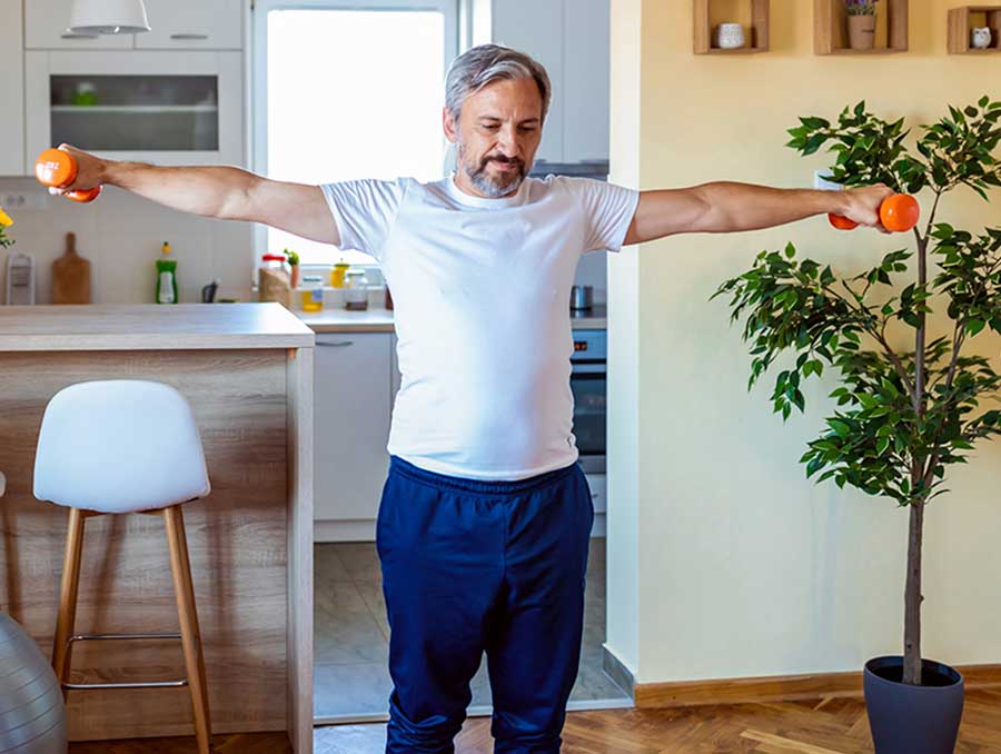
Shoulder Strength and Stability
Wednesday, April 10, 2024 | 6:00-7:00 pm (ET)
2024 National Senior Health & Fitness Day
Wednesday, May 22, 2024 | 10:30-12:00 pm (ET)
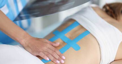
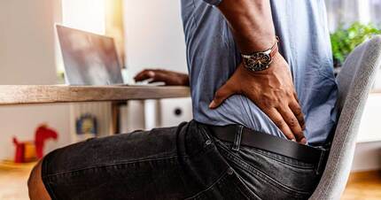
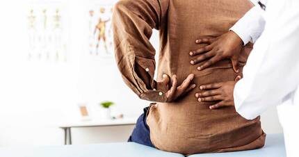
Nearly everyone experiences back pain at some point in their lives. Whether it’s a short-term or more chronic issue, these tips from HSS experts can help you find some relief.

Getting high-quality sleep is just as important as the number of hours you spend snoozing.
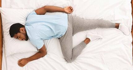
What’s the best sleep position for low back pain? How much does your mattress matter?
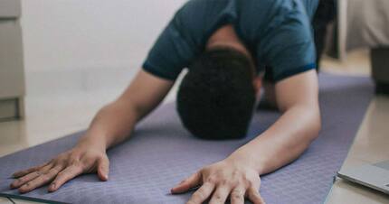
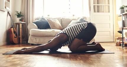
Stretching at night can help you fall asleep faster and avoid sleep-related pain. HSS physical therapists Anna Ribaudo and Sheena Alva have 10 stretches to try.

If you constantly wake up with a stiff, sore neck, your sleeping position may be to blame. Learn how to find relief.
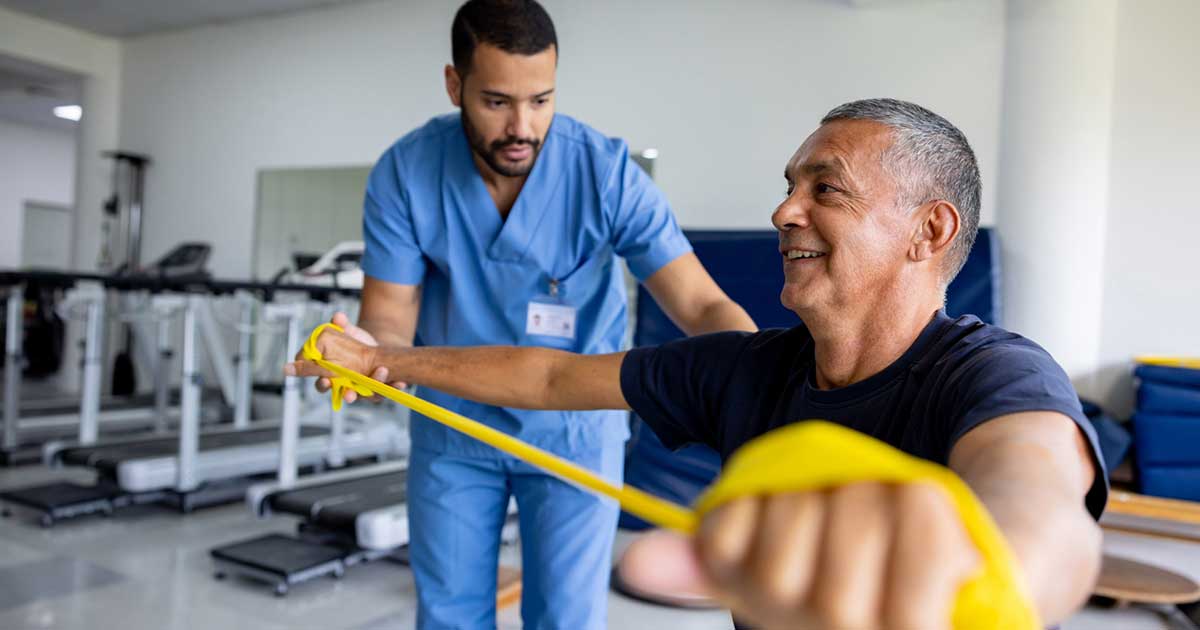
If you’re considering a hip replacement, learn more about how quickly you can get back in action afterward.
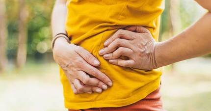
Learn more about this condition that can cause popping noises or sensations in the hip.

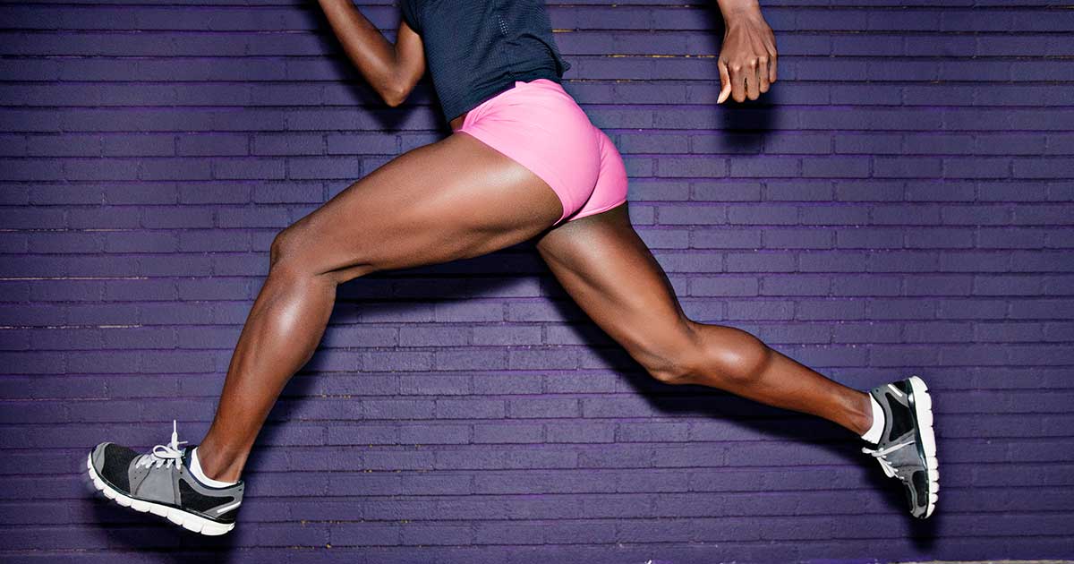
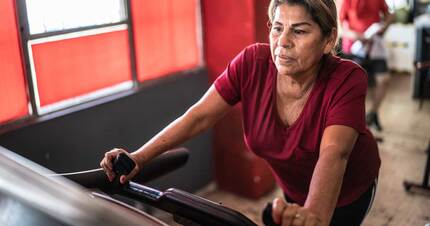
Your hips are an important source of strength, whether you’re powerwalking or strolling the aisles of the grocery store. Here’s how to keep them functioning at their max.