Synovitis Pathology
Synovitis slider
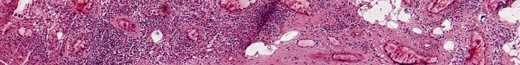
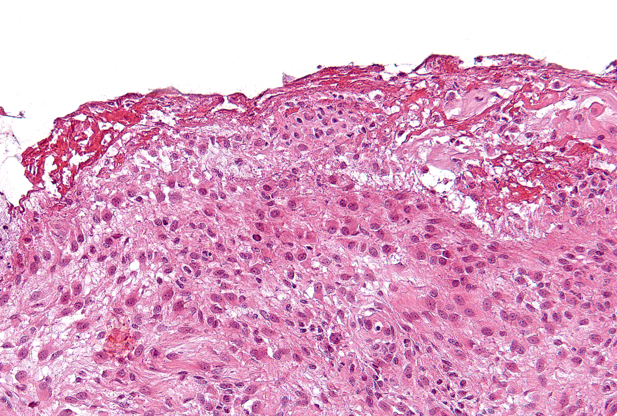
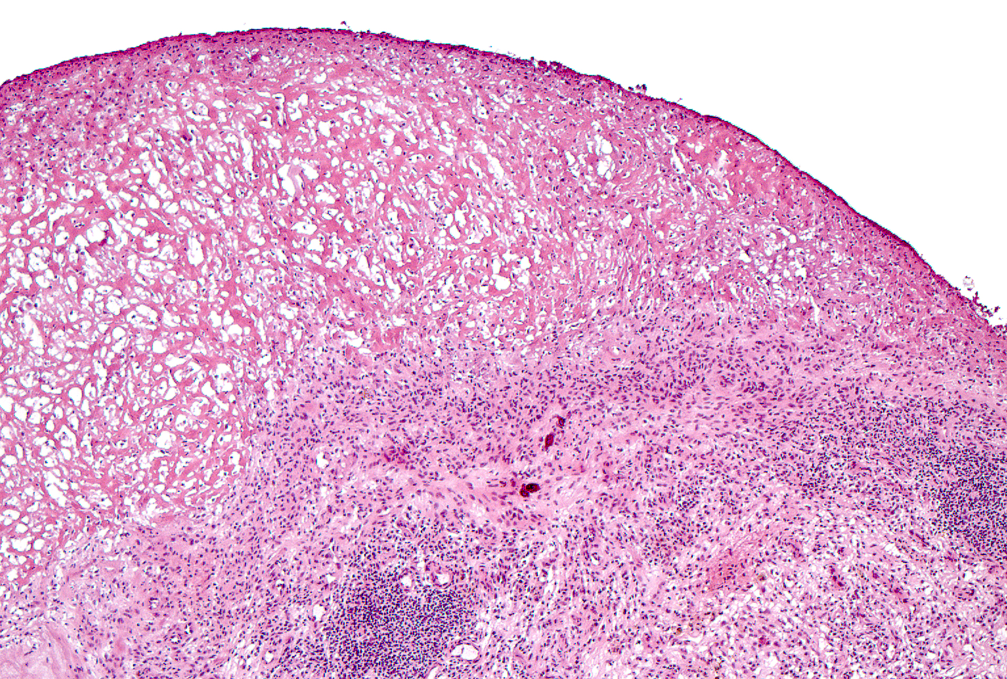
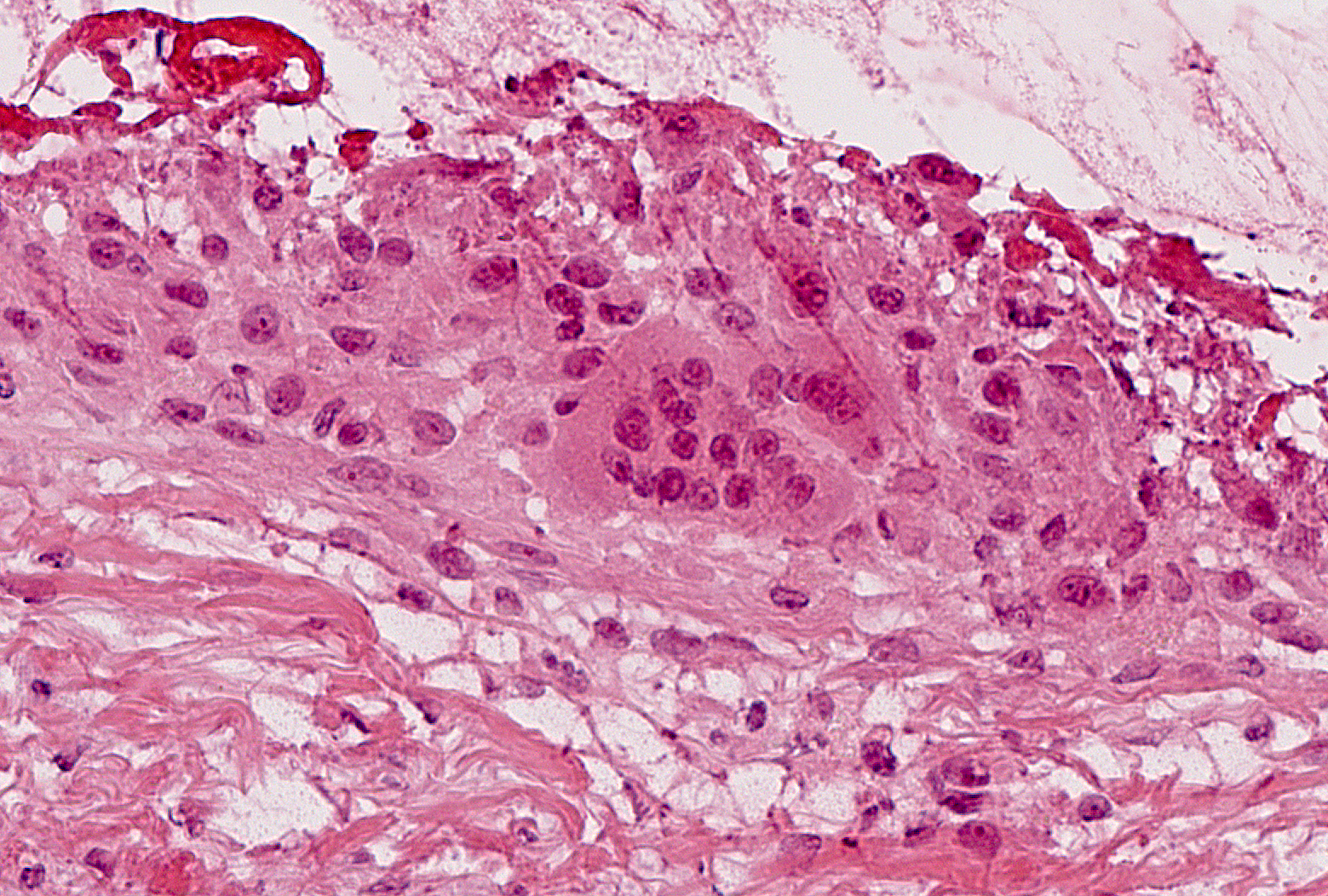
Fibrin and Synovial Giant Cells
Feature:
Fibrillar pink material attached to the surfaces on the synovial membrane
Feature:
Multinucleated giant cells in the synovial lining layer
Fibrin:
Magnification: 4-5x
Grading:
- 0: None (62%)
- 1: Present (38%)
Synovial Giant Cells (SGC):
Magnification: Scan 5x, confirm 20x
Grading:
- 0: None (64%)
- 1: Present (36%)

Back to Pathology of Synovitis