Correction of Bilateral Knee Valgus (Knock Knee) with Osteotomy and Plate Insertion
Patient Case History: Yang
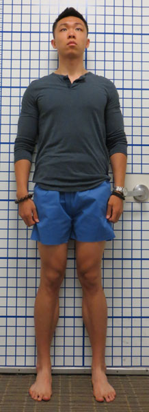
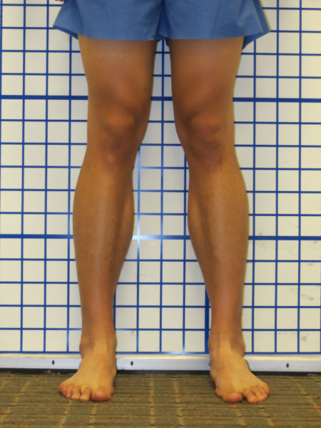
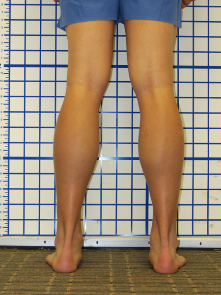
Pre-Op
Patient Surgeon: S. Robert Rozbruch, MD
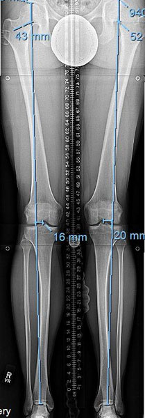
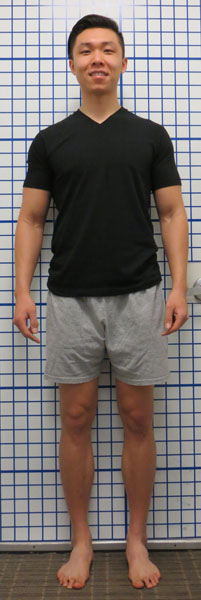
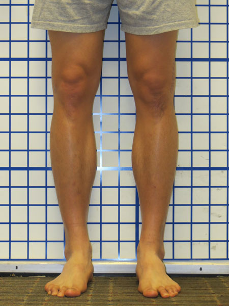
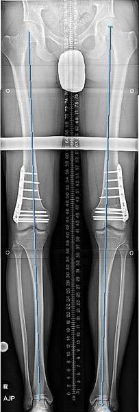
Yang is an athletic young man who was having difficulties related to his knock-knees including pain and awkward gait. He was also concerned with the aesthetics and about further degeneration of the knee in the future. With knock-knees, the lateral joint compartment is overloaded causing pain and progressive degeneration. Notice the line drawn in the x-ray (Figure 4) from hip to ankle passes outside the center of the knee.
Post-Op
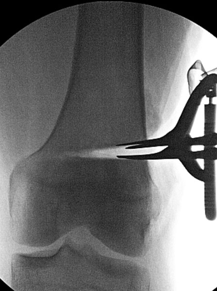
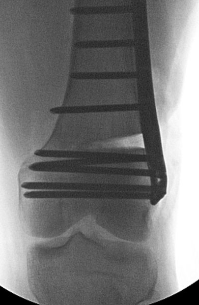
A staged bilateral surgery was performed. The left side was addressed first with an open wedge osteotomy. A partial bone cut was made and the lateral cortex was shimmed open (Figure 1). The alignment was checked in the OR (Figure 2). The new position was secured with a titanium plate (Figure 3) and the space was filled with synthetic bone graft.
Crutches were used for 6 weeks after surgery, and then he began walking normally.

Follow-Up
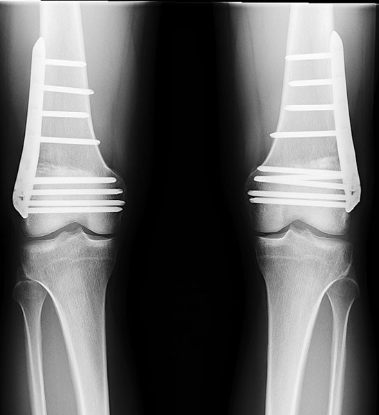
With well healed bones and straight legs, Yang has improved athletically and has no pain. He is very happy with his new appearance and is pleased that the normal alignment helps protect his knees from developing degenerative arthritis in the future.
Notice that the hip to ankle lines pass through the knee center. The two plates were removed one year later during an ambulatory surgery and he was walking the same day.

Return to Patient Case Histories