Click image to close
Previous || Next
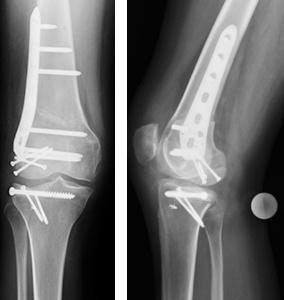
Figure 3. AP and lateral x-rays of the right knee after varus producing distal femoral osteotomy with osteochondral allograft resurfacing of the lateral femoral condyle as well as implantation of an osteochondral hemi-tibial plateau allograft with attached lateral meniscus.