Click image to close
Previous || Next
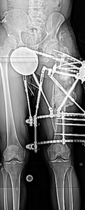
Figure 2: A later standing AP radiograph of both lower extremities showing the Ilizarov/Taylor spatial frame on the left femur. Most of the length is restored and the varus is corrected to a satisfactory position. Note the double rings on the proximal femur used to prevent ring deflection. Also note the bending of the proximal half pins.