Calcaneus Fractures
Case Example
A 47-year-old male fell from a ladder onto his left lower extremity. He was taken to a local hospital and radiographs revealed a left-sided, displaced, intra-articular calcaneus (heel bone) fracture with a depressed articular segment and he was referred to the HSS Orthopedic Trauma Service one day following his injury for definitive treatment. Open reduction and internal fixation (ORIF) was performed by Dr. David L. Helfet and included elevation of the depressed segment, placement of bone graft, fracture reduction, and placement of a calcaneal plate and screws. He returned at regular follow-up intervals and progressive bone healing was noted. At the time of his latest follow-up visit, 5 years following fracture surgery, he has excellent radiographic and clinical results, complete pain resolution, and a return to pre-injury activities.
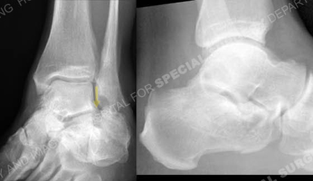
Injury radiographs revealing a calcaneus fracture with a depressed articular segment (arrow).
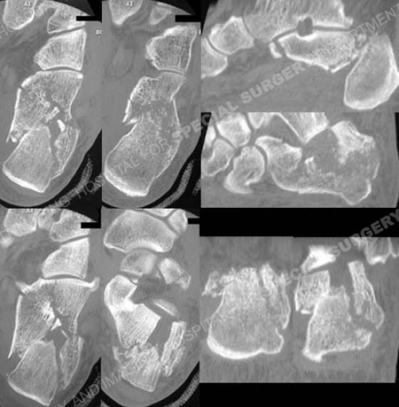
CT scan images further delineating the fracture pattern.
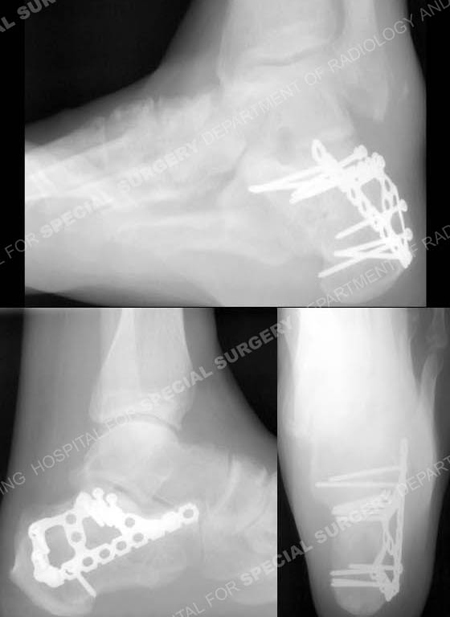
C Broden’s, lateral and Harris heel radiographs (counterclockwise from top) 5 years following fracture surgery
revealing a healed calcaneus fracture in excellent alignment.
Research Publications
The HSS Orthopedic Trauma Service has conducted many studies. Please see our publications on foot fractures.