Minimally Invasive Fracture Surgery
Case Example
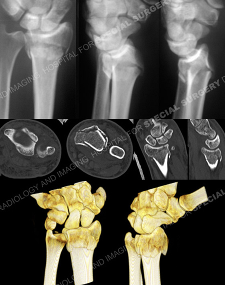
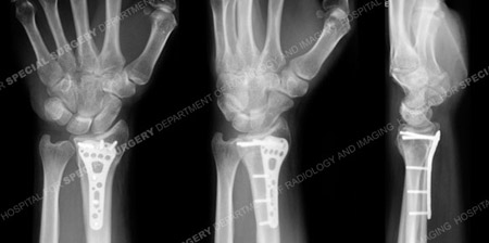
A 34-year-old male fell backwards while snowboarding landing on his outstretched left upper extremity. Radiographs at a local hospital revealed a left-sided unstable distal radius fracture with articular extension and dorsal comminution. A fracture splint was applied and he was referred to Dr. Andrew J. Weiland at the Orthopedic Trauma Service at Hospital for Special Surgery for definitive management. Open Reduction and Internal Fixation (ORIF) was performed with placement of a volar locking plate and screws and indirect reduction of the dorsal comminution. He returned for regular follow-up and healed uneventfully, and at 6 months he presented with excellent results including a healed distal radius fracture in excellent alignment, full range of motion and resolution of pain and returned to all pre-injury activities and sports.

Anteroposterior and lateral injury radiographs (top images) revealing an unstable left-sided distal radius fracture with articular extension and dorsal comminution, loss of volar tilt and radial inclination and height. CT scan and 3D reformatted CT images further delineate the fracture pattern.

Postoperative anteroposterior, mortise and lateral radiographs at 6 months illustrate a healed distal radius fracture in excellent alignment.
Research Publications
The HSS Orthopedic Trauma Service has conducted many studies. Please see our publications on wrist fractures, radius fractures, fractures in athletes, and use of locking plates in fracture treatment.