Ankle Fractures (Broken Ankle)
Broken ankles are painful and temporarily disabling. If a fractured ankle is not properly treated, it can lead to significant, long-term complications and debility.
- What is a broken ankle?
- Anatomy of the ankle joint
- What causes a broken ankle?
- What causes a stress fracture in the ankle?
- What are the different types of ankle fractures?
- What are the symptoms of a broken ankle?
- How is a broken ankle diagnosed?
- How is a broken ankle treated?
- What is the recovery time of a broken ankle?
What is a broken ankle?
A broken ankle is a fracture or multiple fractures of one or more of three bones in the ankle joint: the tibia (shinbone), the fibula (outer bone of the lower leg), and the talus.
Broken Ankle? Get quick access to an HSS orthopedic surgeon.
Call our Ankle Fracture Line at 833.294.9759
Anatomy of the ankle joint
The ankle joint is composed of the tibia, fibula and talus bones. The talus (or "ankle bone") connects your leg to your foot.
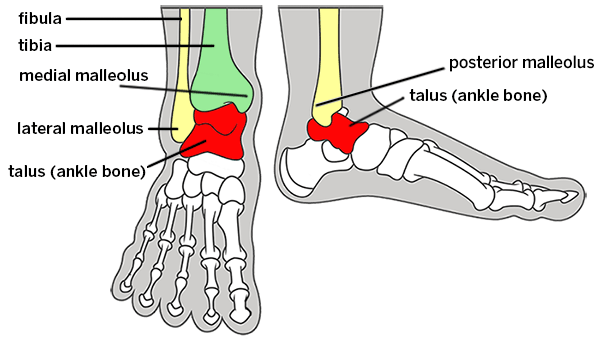
Skeletal anatomy of the ankle
Ligaments connect bone to bone to provide stability of the joints. They are commonly injured in the case of ankle sprains. They can also be injured in connection with ankle fractures. When ligaments are torn and associated with an ankle fracture, this damage can render the ankle unstable. The deltoid ligament is found on the inner part of the ankle and provides the majority of the stability of the ankle. If the deltoid ligament is torn in association with a fracture, the ankle is generally unstable.
In the ankle joint or any joint in the body, two or more bones move relative to one another. There is a cushion or lining between the bones, which is called cartilage. Thinning or damage to this cushion can lead to arthritis or inflammation in the joint.
What causes a broken ankle?
Broken ankles are usually caused by a rotational injury, where the ankle becomes twisted, turned or rolled while walking or running, such as during sports activity. But they can also be caused by a high-force impact, such as from a fall or automobile collision.
Breaks that occur suddenly, during a specific incident or injury, are known as traumatic ankle fractures. But a bone in the ankle can also break due to repetitive stress or impact over time. These are called stress fractures.
What causes a stress fracture in the ankle?
An ankle stress fracture usually occurs some time after a person begins a new activity that involves significant impact of the foot, such as hiking, running or field sports. They can also occur in an active person who quickly increases their activity, for example when someone who is accustomed to jogging a few miles a week begins to train for a 26-mile marathon.
Stress fractures can occur in any of the three ankle bones, especially the tibia or fibula. They are also common in the navicular bone, which is separate from the ankle, but lies directly beneath the talus.
What are the different types of ankle fractures?
Because the ankle joint comprises three bones, there are numerous types of ankle fractures. Doctors think of the ankle as having three sides and a "roof," and fractures can occur in each of these areas or in combination.
The lower portion of the tibia forms the roof and medial (inside) of the ankle, while the lower portion of the fibula forms the lateral (outside) and posterior (back) of the ankle.
Most common ankle fractures
- Lateral malleolus fracture: This is the most common type of ankle fracture. It is a break of the lateral malleolus, the knobby bump on the outside of the ankle (in the lower portion of the fibula).
- Bimalleolar ankle fracture: This second-most common type involves breaks of both the lateral malleolus and of the medial malleolus, the knobby bump on the inside of the ankle (in the lower portion of the tibia).
- Trimalleolar ankle fracture: This type involves breaks in three sides of the ankle: the medial malleolus, the lateral malleolus and distal (lower portion) of the posterior malleolus of the tibia.
- Pilon fracture (also called a plafond fracture): This is a fracture through the weightbearing “roof” of the ankle (the central portion of the lower tibia). This is usually a higher energy traumatic injury resulting from a fall from a height.
As the number of fracture lines increase, so does the risk of long-term joint damage. Trimalleolar ankle fractures and pilon fractures have the most cartilage injury and, therefore, have a higher risk of arthritis in the future.
Nondisplaced vs. displaced ankle fractures
Within each of the above types, the fracture will be either:
- Nondisplaced – Bones are broken but still in correct position and alignment.
- Displaced – Fractured portions of bone are separated or misaligned. The treatment will be based on fracture alignment and stability of the ankle.
There are some additional, unique types of fractures.
Maisonneuve fracture
A Maisonneuve fracture, for example, involves a complete disruption of the ligaments around the ankle associated with a fracture of the fibula at the level of the knee. For this type of injury, an ankle X-ray may not show a fracture or demonstrate the instability associated with this injury, because the actual bone fracture is well above the ankle, and the ligament injuries can only be seen with other forms of imaging, such as an MRI.
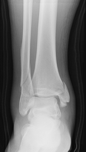
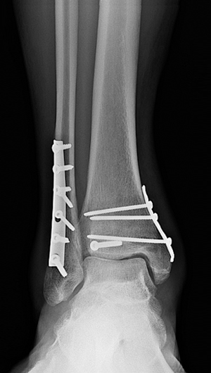
X-ray image showing front view of a bimalleolar fracture
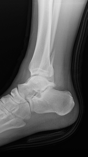
X-ray image showing side view of a displaced lateral malleolus fracture of the right ankle
What are the symptoms of a broken ankle?
The most common symptoms of an ankle fracture are pain and swelling, either of which may be present only in the ankle region itself or spread to parts of the foot or up toward the knee. Any pain will usually be more intense if the injured person tries to put weight on the ankle.
How is a broken ankle diagnosed?
X-rays are usually required to determine whether there is a broken bone as opposed to a soft-tissue injury like a sprain, since ankle sprains and breaks have similar symptoms. Other radiology imaging, such as a CT scan or MRI, may be needed to determine the full scope of the injury.
If the imaging shows that a person has a fractured ankle, he or she should consult an orthopedic surgeon as soon as possible. There are several different types of ankle breaks, and not all require surgery. But when they do, it is important that they receive the appropriate surgery by a skilled foot and ankle specialist. An inappropriate or poorly executed surgery can lead a patient to require additional corrective surgeries and/or, years later, to develop ankle instability, arthritis or even the need for an ankle replacement operation. Early and correct intervention is the key to preserving the ankle joint over the long term.
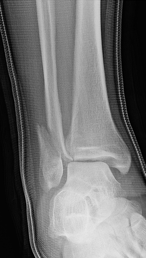
X-ray showing front view of a displaced fibula with medial clear space widening (asymmetry of the joint space) indicating a deltoid ligament disruption
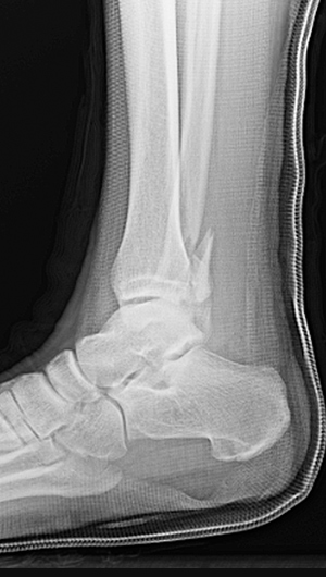
X-ray showing side view of a displaced fibula fracture with a posterior malleolar fracture
How is a broken ankle treated?
Treatment is based on the alignment of the bones and the stability of the ankle joint. The goal is to have the bones heal as closely to perfect as possible so as to prevent any residual instability or malalignment of the bone. A malalignment of as little as two millimeters in the ankle joint can lead to arthritis. It is much easier to fix a fracture than to treat arthritis in the future. Certain mild ankle breaks (stable and with no displacement) can be treated nonsurgically with a splint, short leg cast, or other protective device such as a walking boot Some patients may be able to walk immediately while wearing a support while others may have to use crutches to limit weightbearing.
For more serious fractures in which bones or bone fragments are misaligned, surgical intervention is necessary to prevent improper healing (malunion) that would impede proper movement in the ankle and possibly lead to other complications.
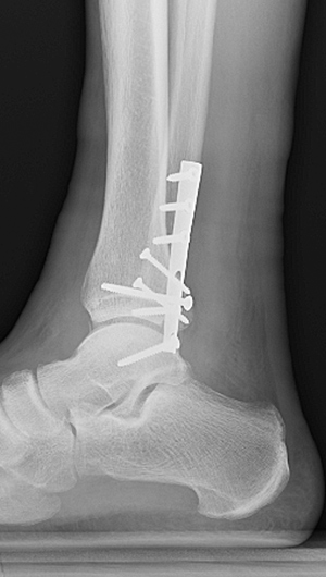
X-ray showing front view of surgical plates and screws to treat a trimalleolar fracture
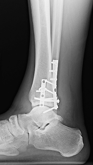
X-ray showing side view of the same
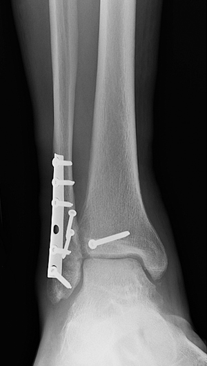
X-ray showing front view of fixation of the fibula and posterior malleolus with restoration of the joint congruity
X-ray showing side view of the same
What is the recovery time of a broken ankle?
It takes about six weeks for bones to heal. It may take longer for ligaments or other soft tissues to heal as well.
After surgery, patients are typically not weightbearing for 4 to 6 weeks until the bone heals. Patients are placed on a pain management protocol that minimizes their need for opioid medications. For the first couple of weeks, patients are in a splint and are elevating the limb 90% of the day. After 10 to 14 days, the sutures are removed and patients are typically placed into a removable boot. This allows patients to start moving the ankle and to shower. At the six-week visit, X-rays are obtained. Assuming the bone is healed well, patients are then allowed to start weightbearing and to begin physical therapy. Patients will generally have six weeks of therapy or more if required.
Ankle fracture articles for patients
Read the below articles for discussions on pediatric ankle fractures and stress fractures.
Ankle Fractures (Broken Ankle) Success Stories
In the news