Pediatric Foot Deformities
- Pediatric Foot Deformity: An Overview
- Cavus Foot
- Tarsal Coalition
- Clubfoot
- Accessory Navicular
- Juvenile Bunion
- Finding the Right Treatment for Pediatric Foot Deformity
Pediatric Foot Deformity: An Overview
Pediatric foot deformity is a term that includes a range of conditions that may affect the bones, tendons, and muscles of the foot. Among those most frequently treated at HSS are cavus foot, tarsal coalition, clubfoot, accessory navicular, and juvenile bunion.
Treatment of foot deformities in children can vary significantly from that needed in adults. Fortunately, pediatric orthopedists who specialize in this field can bring to bear a range of non-operative and operative techniques specifically developed to address the distinctive needs of children, which include special attention to preserving the integrity of the growth plate, allowing continued growth and development of the foot.
Cavus Foot
Cavus foot is a condition in which the child has an excessively high arch. In many cases, the heel of the foot is turned inward (this is known as cavovarus foot deformity). The condition frequently affects both feet, and it is often progressive. Patients experience pain and develop calluses because is not properly aligned, which causes uneven weight-bearing. Ankle sprains or even stress fractures may occur.
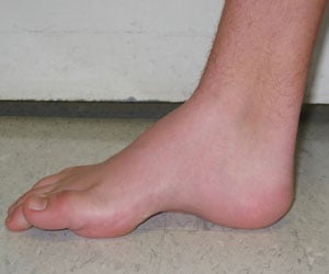
Cavovarus foot – Medial to lateral view of a right
cavovarus foot. Note the pathognomonic high arch.
A cavovarus foot that develops over time (as opposed to that which can appear with club foot; see below) can indicate the presence of a neurologic problem, the presence of a cyst, or possibly a tumor in the spinal cord.
More commonly, however, it is the result of Charcot-Marie Tooth disease, a hereditary disorder in which the conduction speed of nerves slow over time and causes weakness of the distal muscles of the hands and feet. While Charcot-Marie Tooth disease is not life-threatening, the foot problems that accompany it can be disabling. In fact, because other symptoms may be mild or non-existent, the cavovarus foot may be the key to diagnosis.
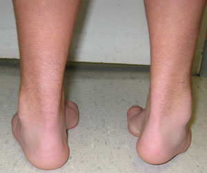
Posterior view of the same patient showing cavovarus foot in both feet.
In cases of mild cavus deformity, the orthopedist may recommend use of an orthotic (a shoe insert that can help evenly distribute the way in which the foot bears weight) and monitoring for progression of the problem. Owing to the need to realign the foot, however, cavus deformity often requires surgical treatment, consisting of a combination of osteotomies, in which the bones are cut and repositioned, ligaments are released and tendons are transferred to help rebalance and supplement weakened muscles in the foot and leg.
“Correcting cavus deformities is complex,” explains Dr. David M. Scher, Associate Attending Orthopedic Surgeon at HSS. “There is no ‘cookbook’ for deciding exactly what surgeries to do, as each child is different.” Among the factors the surgeon must consider is the appearance of the foot, information obtained by X-ray, the physical exam and patient symptoms.
Growth potential must also be taken into account. “How we cut the bones, and the ways in which we put in the pins and screws during surgery is different in children than it would be in the adult since the foot must be allowed to grow.” Dr. Scher also emphasizes the importance of seeking treatment from a pediatric orthopedist, cautioning that although adult foot and ankle surgeons may have extensive experience in treating this condition, the special needs of children require a different approach.
Tarsal Coalition
Children with tarsal coalition develop an abnormal connection between the bones in the midsection and back part of the foot. It is usually diagnosed in late childhood or early adolescence when the coalition begins to limit foot movement, causing pain and sometimes stiffness. Symptoms may be particularly noticeable when walking on an uneven surface, such as sand or gravel, an action that requires constant adjustment of the foot. Frequent ankle sprains may also signal the presence of a coalition.
Most tarsal coalitions may be classified as one of two types: a calcaneonavicular coalition, in which the tissue develops between the calcaneus (heel bone) and the navicular (one of the foot bones), or a talocalcaneal or subtalar coalition, in which the coalition develops between the calcaneus and the talus (the ankle bone). The coalition may be composed exclusively of bone, a combination of bone and cartilage, or even fibrous tissue. Tarsal coalitions occur in both feet in about half of all cases.
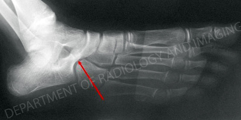
Right foot with calcaneonavicular coalition. The red arrow indicates the coalition.
Initial treatment for tarsal coalitions is non-operative; the patient is required to rest or immobilize the foot. Although pain relief can be achieved in this way, in many cases the result is only temporary. Non-operative treatment can be appropriate for patients with tarsal coalitions that are symptom-free, and whose condition only becomes apparent on x-ray taken incidentally for another condition, such as an acute ankle sprain.
For children with ongoing pain, who do not respond to non-operative treatment, a pediatric orthopedist may perform a resection or removal of the coalition tissue. In most cases, in order to prevent regrowth of the coalition, another type of tissue – usually fat – is placed between the bones, to prevent the coalition from growing back.
"This is a fairly simple operation with the child typically back on his or her feet in between 2 to 6 weeks," says Scher. Occasionally, children with tarsal coalitions may have another type of foot deformity as well, such as rigid flatfoot, which can be painful. If this deformity is severe, additional surgery may be required to realign the foot.
Clubfoot
Clubfoot is a condition in which the foot – or sometimes both feet – are turned inward and are pointing down. Immediately apparent at birth, clubfoot is known to develop during intrauterine life, at between 9 and 14 weeks gestation. In fact, in many cases, the deformity is detected on routine ultrasound. “This can make an enormous difference for parents,” says Dr. Scher. “It’s much easier for parents to cope and plan when they know in advance what they have to do.”
Occurring in about one in 1000 births, clubfoot is statistically seen more frequently in boys than in girls. And although family history may play a part, many infants with clubfoot have no known relative with the condition.
In the majority of cases, clubfoot can be successfully treated without surgery, using the Ponseti technique, which employs gentle manipulation and casting of the feet at weekly intervals. Treatment begins shortly after birth when the newborn’s foot, including tendons, ligaments, joint capsules and bones are most responsive. Following this first phase of treatment, a brace is used for an extended period to maintain proper alignment. When applied correctly, the Ponseti technique yields excellent results.
In some instances, clubfoot does require surgery – the approach that was used historically, before widespread acceptance of the Ponseti technique. While this approach can yield good correction of the deformity, the procedure may result in a stiff and arthritic foot later in life. When this occurs, additional surgeries may be necessary to alleviate the arthritic pain.
Accessory Navicular
Accessory navicular describes the presence of an extra bone growth center on the inside of the navicular and within the posterial tibial tendon that attaches to the navicular. The primary symptom from this additional bony prominence is pain and tenderness.

X-ray image of the left foot with an accessory navicular.

Enlarged view of the affected area. The red arrow indicates the accessory navicular.
This congenital defect (present at birth) is thought to occur during development when the bone is calcifying. Because this accessory portion of the bone and the navicular never quite grow together, it is believed that, over time, the excessive motion between the two bones results in pain.
The initial treatment approach for accessory navicular is non-operative. An orthotic may be recommended or the patient may undergo a brief period of casting to rest the foot. For chronic pain, however, the orthopedic surgeon removes the extra bone, a relatively simple surgery with a brief rehabilitation period and a very good success rate.
Juvenile Bunion
As with bunions in adults, in juvenile bunion, the joint at the base of the big toe (the metatarsophalangeal joint) moves out of alignment in such a way that the big toe angles inward to the second toe.
However, unlike adult bunion, which usually results from ill-fitting footwear or has a hereditary component, juvenile bunion occurs most often in children who are ligamentously lax or loose-jointed. The problem is more common in girls than in boys.
Surgical treatment for juvenile bunion is generally restricted until the end – or close to the end – of growth, both because of concern for damage to the growth plate and because the condition tends to recur. Non-operative treatment includes the use of wide shoes or sneakers and avoidance of narrow dress shoes and high heels. Usually this sufficiently alleviates symptoms to avoid or defer the need for surgery.
In younger patients who do not respond to non-operative treatment and who have pain that interferes with their daily activities, surgery to realign the bone and straighten the toe can be performed. A number of different approaches are used, depending on the type of bunion, the extent of the deformity, the age of the child, and how much growth remains.
Finding the Right Treatment for Pediatric Foot Deformity
For parents seeking treatment for children with foot deformities, Dr. Scher advises consultation with a pediatric orthopedic surgeon with extensive experience in his or her field. Not only will they be aware of all the relevant non-operative techniques, but should surgery be necessary, “overall, the more procedures the surgeon does, the better the success rate,” he says. Centers like Hospital for Special Surgery also offer patients and their families the benefits of general pediatricians on staff as well as anesthesiologists and physical therapists who specialize in pediatric care.
For more information on treatment of pediatric foot deformities at HSS, please call our Physician Referral Service at 1.877.606.1555.