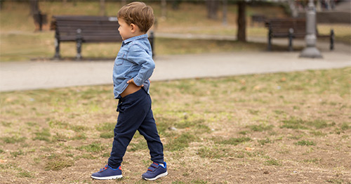
Legg-Calve-Perthes Disease: An Overview
Legg-Calve-Perthes disease (also called Perthes disease) is an uncommon pediatric hip disorder. It may cause a painless (or occasionally painful), intermittent limping and limited hip motion in growing children.
What is Legg-Calve-Perthes disease?
Legg-Calve-Perthes disease, also known as Perthes disease, is a rare disorder of the uppermost part of the femur bone, called the femoral head. The femoral head is the ball of the ball and socket joint of the hip. There is a disruption of normal blood flow to the femoral head and the bone suffers an ischemic attack. The femoral head then goes through a series of stages over a one- to three-year period.
First the bone dies, next it fragments, then it regenerates, and finally it heals as the blood supply returns to the femoral head. This process affects the growth and development of the hip joint. There is variability in how much the hip joint is affected based on the child’s age of onset of Perthes as well as the extent of initial ischemia to the femoral head.
Perthes disease affects boys five times more than girls between 3 and 10 years of age. In about 12% of cases, it may affect both hips asynchronously.
What are the symptoms of Legg-Calve-Perthes disease?
Legg-Calve-Perthes disease, also known as Perthes disease, is one of many possible causes of hip pain in children 3 to 10 years old. A child with Legg-Calve-Perthes disease will limp and sometimes complain of thigh or knee pain. Persistent limping as well as limited motion of the affected hip are what generally lead to a doctor’s evaluation and eventual referral to a pediatric orthopedic surgeon.
If a limp is fleeting for one or two days and never returns (unless a new injury occurs during play, for instance, causes pain), there should be no concern. However, if a limp is steady or intermittent and persists or recurs, it is appropriate to consult a pediatric orthopedic surgeon.
What causes Legg-Calve-Perthes disease?
It is widely believed that Perthes is caused by idiopathic avascular necrosis of the femoral head. That is, arising spontaneously from an idiopathic (unknown) cause, the top section of the femur (thighbone), known as the femoral head, which inserts into a socket of the pelvic bone to form the hip joint, dies due to a lack of blood supply (avascular necrosis).
Neither scientists nor physicians understand why the blood supply to the femoral head becomes compromised in such cases. Attention deficit disorder and coagulopathies have been associated with Perthes. The bone of the femoral head collapses and then regrows and remodels. The entire process takes approximately one to three years.
During that time, the pediatric orthopedic surgeon will monitor the child through regular follow-up appointments every four to six months to check hip range of motion, gait (walking pattern) and pelvic radiographs (X-rays).
How is Legg-Calve-Perthes disease diagnosed?
Perthes is diagnosed with a clear history of what is happening from the parent and child, a physical examination of a child’s gait and lower extremities, and a radiograph of the pelvis. If the pediatric orthopedic surgeon suspects Perthes based on the history and physical examination, but it is not clear on the pelvic radiograph, and MRI of the hip is a helpful tool to help detect Perthes in the early stage.
How is Legg-Calve-Perthes disease treated?
Depending on the findings that result from the child’s physical exam and X-rays, treatment will range from nonsteroidal anti-inflammatory medications (NSAIDs), such as ibuprofen, and activity modification (avoiding running and jumping activities) to physical therapy. Sometimes braces are utilized and rarely surgery to redirect the orientation of the femoral head to optimize the congruity of the hip joint. Orthopedic surgery, though rarely required, may be performed to enhance hip joint congruity when necessary.
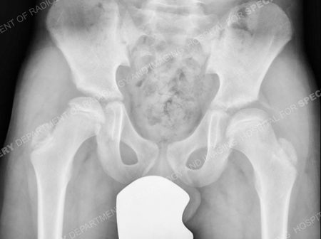
Figure 1: Typical x-ray appearance of an 11-year-old child in the early stages of Perthes disease
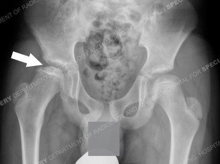
Figure 2: Eight months later, the femoral head is collapsing and fragmenting (as depicted by the white arrow).
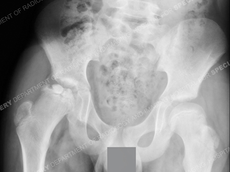
Figure 3: Ten months after the first x-ray (Figure 1), the femoral head is very irregular at this point and is subluxating (coming out of the socket). Surgery is recommended to redirect the femoral head into the socket (acetabulum) to promote the best chance of healing.
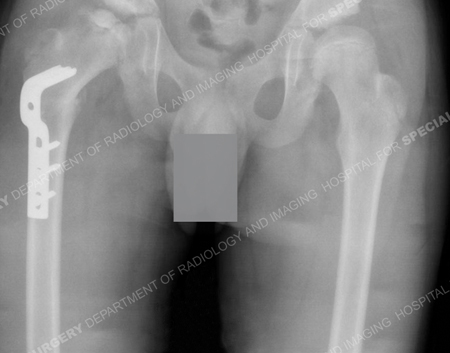
Figure 4: Surgery has been performed. The femoral has been cut, a metal plate is attached, and the femoral head is redirected into the acetabulum.
Does Legg-Calve-Perthes disease affect height?
Perthes changes the shape of the top of the femur which often results in a slightly shorter and broader femoral head. This usually results in a insignificant and asymptomatic leg length inequality, but the pediatric orthopedic surgeon will monitor leg lengths for extreme cases where an intervention may be needed to equalize the length of lower extremities.
What is the long-term outlook for children with Legg-Calve-Perthes disease?
Children with Perthes need to be mindful of their physical activities. Participation in preservation activities, such as swimming or biking, rather than running and jumping sports, will prolong the possible need of hip replacement surgery as an adult.
Physical therapists play an important role in the care of patients with Perthes. The physical therapist will work with the child on range of motion and strengthening exercises for the lower extremities, as maintaining strength of the lower extremities and range of motion of the affected hip is very important in the rehabilitation of a child with Perthes disease.
Updated: 7/25/2023
Authors
Associate Attending Orthopedic Surgeon, Hospital for Special Surgery
Associate Professor of Orthopedic Surgery, Weill Cornell Medical College
