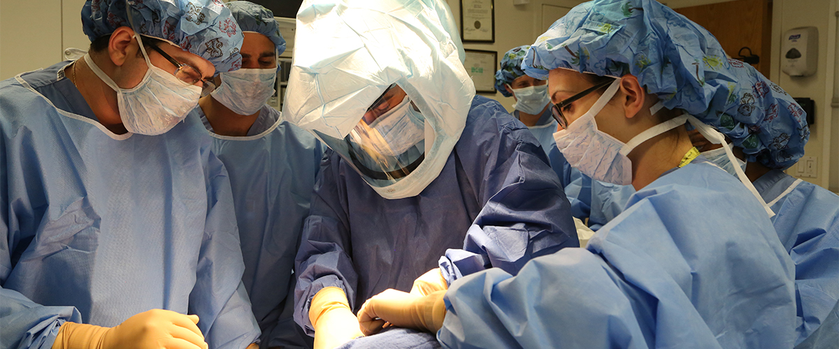
Cadaver Lab slider
Cadaver Lab Curriculum
Anatomy: The Sine Qua Non of Regional Anesthesia
In 1884, Dr. William Halstead performed what is believed to be the first peripheral nerve block. That Dr. Halstead was a surgeon should come as no surprise — the anesthetic technique he employed consisted of neck dissection followed by direct injection of local anesthetic into the nerve roots of the brachial plexus.
By today’s standards, his bold approach to conduction blockade seems unnecessarily invasive. At the time, however, surgical exploration remained the sole means by which to locate the site of injection.
Today, anesthesiologists have a variety of percutaneous nerve localization techniques at their disposal. Mechanical parasthesia, peripheral nerve stimulation, and lately, ultrasound guidance allow for far less invasive access to the target nerves. While the days of surgical exploration are long gone, knowledge of anatomy and its clinical application has remained the cornerstone of all peripheral nerve practice.
Educating the Peripheral Nerve Block Practitioner
Primum non nocere, or "first do no harm", mandates that peripheral nerve block practitioners achieve and maintain a detailed understanding of the relevant anatomy and common pitfalls associated with each block technique.
In the past, such knowledge was acquired through independent review of available anatomic literature. Progress in regional anesthetic techniques was therefore slow and isolated.
Over the last twenty or so years, much has changed. A handful of regional anesthesia centers, HSS among them, have greatly aided in the dissemination of knowledge pertinent to regional anesthesia through residency and fellowship training programs.
Still, the path to nerve block competency has remained challenging – primarily because students of the specialty have lacked access to a suitable model on which to practice and perfect a given block technique. Traditionally, patients have filled this role.
Under the watchful eye and steady hand of mentoring anesthesiologists, residents and fellows cautiously acquired the practical knowledge of peripheral nerve blockade. While this educational model has proved safe and successful, it has invariably limited the rate at which the student attains mastery of a given block.
A Novel Way to Teach Regional Anesthesia
Unlike typical cadaver courses which make use of embalmed specimens, the lab employs fresh frozen cadavers which replicate the look and feel of living tissue. Because these specimens are not embalmed, ultrasonography yields images that rival those found in the operating room while needle placement and local anesthetic injection mimics the proprioceptive feedback of living tissue.
Students in the lab have the opportunity not only to dissect and discern the anatomy relevant to a particular nerve block but also practice block and catheter placement techniques on “patients” in an environment free of time constraints or fear of complications.
Highlights of the curriculum include:
- A review of various anatomic approaches to each block
- An examination of the disparate techniques of nerve localization including mechanical parasthesia, nerve stimulation, and ultrasound guidance
- A focus on the proprioceptive feedback of distinct tissues including blood vessels, bone, muscle, nerve, and lung
- The use of methylene blue injectate to map the distribution of local anesthetic following injection
- The study of complications including intravascular, intraneural, and transforaminal injection, as well as pneumothorax and its tell-tale signs
In all, our cadaver lab provides students with regional anesthesia the substrate and time necessary to develop the knowledge, skill, and proprioception critical to safe, efficacious peripheral nerve blockade.
eAcademy Cadaver Lab Courses
- Blocks for the Upper Extremity: Interscalene and Supraclavicular
- Slide Series: Interscalene Block
- Slide Series: Sciatic Nerve Block
- Slide Series: Adductor Canal Block
Back to Regional Anesthesiology & Acute Pain Medicine Fellowship